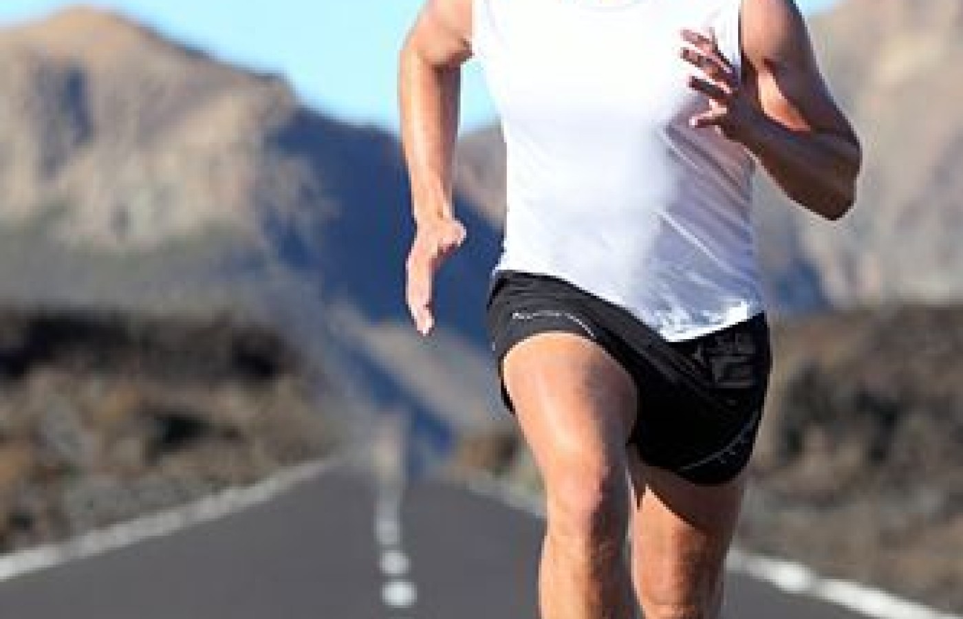
Stress injuries to bony tissues are common occurrences, especially among active individuals.1 Chiropractors are often consulted for the vague pain and nonspecific symptoms of early stress fractures. These injuries respond very well to chiropractic management, especially when postural support is included.
Thanks to advanced imaging procedures, we now better understand that stress fractures result from the inability of bone to withstand excessive and repetitive microtraumas, which in themselves would not be sufficient to cause a fracture.2 Overuse syndromes such as stress fractures are due to biomechanical stress that exceeds the body's inherent capacity to repair and adapt.3 In other words, the repair process cannot keep up with the stressor and is overcome, with the eventual result that the tissue fails, resulting in injury. These conditions must be managed properly by the treating chiropractor, since any delay in establishing an appropriate treatment plan will result in continuing progression and true fracture of bone, placing the patient at risk for a disabling condition.4-5
A Quartet of Microtrauma Sources
Four major sources of microtrauma can result in stress reactions: overuse, poor training techniques, inherent biomechanical stresses, and poor shock absorption. Overuse occurs when a body part is exposed to unusual, repetitive trauma such as starting a new running program. In this case, everything is done correctly, but there is just too much stress, too soon.
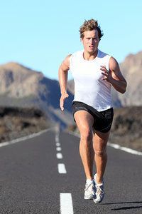
Poor training techniques include running long mileage on one side of the road, resulting in excessive stress due to the angulation of the road. Training techniques can usually be easily changed to reduce physical stress.
Biomechanical stresses are occasionally high due to inherent imbalances or asymmetries, which often require modification of performance or equipment for complete resolution. And poor shock absorption can result from running on unyielding surfaces or in broken-down shoes, or may be due to inherent factors which interfere with normal shock absorption, such as a high-arched foot.
Since up to 95 percent of stress fractures occur in the lower extremities (including the hip),6 most incidents of stress reaction in bone seen by chiropractors will localize to the foot, leg, hip or pelvis. Typically the patient will report a dull, aching pain sensation that is poorly localized. The nagging pain will increase during use of the affected area, and often remain for a while after use; then subside or disappear with rest overnight.
Involvement of the femoral neck will often cause aching discomfort to radiate into the groin and posterior pelvis.5 The most important aspect of the symptom pattern for accurate diagnosis is the recurrence of pain with exercise and the relief of pain with rest.
A common source of excessive biomechanical stress to bones of the lower extremity is malalignment or asymmetry. During standing, walking and running, the lower extremities and pelvis form a closed kinetic chain. Torque, shock, tension and compression forces are then transmitted between the bones, joints and muscles. If imbalances or asymmetries are present (whether developmental or acquired), these forces can reach excessive levels and eventually cause or contribute to a stress fracture.
A Trio of Conditions
Three specific conditions have been studied that can be easily noted and corrected in the chiropractic office: the high arch, excessive pronation (with a low arch), and inequality of leg lengths.
High arch. The foot with a higher-than-normal arch (cavus foot) remains too rigid and inflexible during walking and running. This results in poor attenuation of heel-strike shock, much of which is then transmitted up the kinetic chain into the leg and hip. This type of foot requires better flexibility and individually designed orthotics with added shock-absorption material to dynamically support the arches and help decrease shock at heel strike.7
Excessive pronation (rolling inward of the ankle) during walking and/or running may be due to either arch collapse or poor arch development. In either case, excessive torsional (twisting) forces are transmitted from the overpronated foot into the leg with each step taken.8 Orthotic support for the arches that includes pronation correction at the heel (a medial or "varus" wedge) will decrease the torque forces on the foot and leg bones and prevent the development of stress reactions.9
Leg-length discrepancy is another inherent asymmetry that has been shown to lead to increased frequency of stress fractures. Several studies9-10 have found that degenerative changes in the hip joint and lumbar spine occur much more frequently on the side of a longer leg. Treatment consisting of a flexible orthotic and/or shoe build-up to reduce the discrepancy is appropriate, in order to decrease the abnormal biomechanical forces developed during walking and running with asymmetrical leg lengths.11
Changing / Moderating Activity
Rest of the patient's affected extremity is necessary initially in order to allow for healing and remodeling.
Bone healing does not demand complete bed rest, but rather a change and moderation of activity. Temporary cessation of the causative exercise – with substitution of cross-training – will prevent deconditioning of the athlete. Low-stress exercise programs that utilize controlled isotonic protocols (such as exercise tubing) can provide benefits from the beginning of healing through to the achievement of previous levels of activity.
References
Lempainen L, et al. Medial malleolar stress fracture in athletes: diagnosis and operative treatment. Scan J Surg, 2012;101(4):261-264.
Yochum TR, Rowe LJ. Essentials of Skeletal Radiology, 2nd Edition. Baltimore: Williams & Wilkins, 1996:460.
O'Conner FG. Overuse injuries in athletes. Phys Sports Med, 1992;20(10):128-142.
Johansson CI, et al. Stress fractures of the femoral neck in athletes: the consequence of a delayed diagnosis. Am J Sports Med, 1990;18:524-528.
Lemire JJ, O'Conner SM. Femoral neck stress fracture, a potentially disabling condition: a case report. J Can Chirop Assoc, 1993;37(2):85-91.
McBryde AM. Stress fractures in runners. Clin Sports Med, 1985;4:737-752.
Simkin A, et al. Combined effect of foot arch structure and an orthotic device on stress fractures. Foot & Ankle, 1989;10(1):25-29.
Michaud TC. Recurrent lower tibial stress fracture in a long-distance runner: a case report. Chirop Sports Med, 1988;2(3):78-87.
Radin EL, et al. Role of mechanical factors in pathogenesis of primary osteoarthritis. Lancet, 1973;519-522.
Giles LGF, Taylor JR. Lumbar spine structural changes associated with leg length inequality. Spine, 1982; 7(2):159-162.
McCaw ST, Bates BT. Biomechanical implications of mild leg length inequality. Br J Sports Med, 1991; 25(1):10-13.
Many relevant diagnostic signs are not performed deliberately by the examiner or by the patient at the examiner’s direction. They are observed as the patient reacts to their condition. Fortin’s finger sign, Minor’s sign, and Vanzetti’s sign are three examples of this principle.
Spearheaded by burgeoning scientific and clinical research literature, psychedelics have reached a level of media coverage and popular interest that has not been seen for over half a century. By “psychedelics,” we are referring to the unique class of substances that includes psilocybin (the active compound found in so-called “magic mushrooms”), LSD, dimethyltryptamine (DMT), ayahuasca, 5-MeO-DMT, and mescaline – each of which occurs in the natural world (except for LSD, which is a semi-synthetic compound).
While working at the Lovelace Medical Center in Albuquerque, N.M., one of the primary care physicians stated that imaging every patient prior to receiving chiropractic was not medically necessary. It was at that point I reconsidered the need for the imaging of every patient to avoid malpractice.