Are You Evaluating Your Patients for Osteoporosis?
Deborah Pate, DC, DACBR
The National Osteoporosis Foundation (NOF) recommends drug therapy for osteoporosis in patients with T-scores of -1.5 or lower and other risk factors. The NOF also recommends drug therapy for patients with T-scores of -2.0 or lower and no other risk factors. No, I am not referring to baseball scores; this regards osteoporosis, which affects approximately 28 million people in the United States.2 I know I have addressed this topic before, but I am now much more aware of this disorder. In fact, this evening, I read about 20 studies on osteoporosis, and noted three compression fractures in three different patients - all men, aged 23, 56, and 83 years. In general, I don't expect to see many compression fractures in that small a series of studies, so I guess I was impressed.
I wonder how many patients have osteoporosis that we don't know about until it is too late and they have compression or hip fractures. By that time, the prognosis for recovery is long, and for many, the quality of normal activities of daily living has already begun to diminish. These days, osteoporosis is too common, even in young people, to be ignored. The diagnosis of osteoporosis should be made before the patient suffers a compression fracture or hip fracture. It can be avoided.
Dual-energy X-ray absorptiometry (DXA) is a relatively inexpensive measurement for bone density. DXA provides information about bone density and fracture risk. Diagnosis is based on calculated T-scores:
BMDindividual - BMDyoung normal
BMDyoung normal
Normal: > -1.0 Osteopenic: -1.5 to -2.5 Osteoporotic: > -2.5
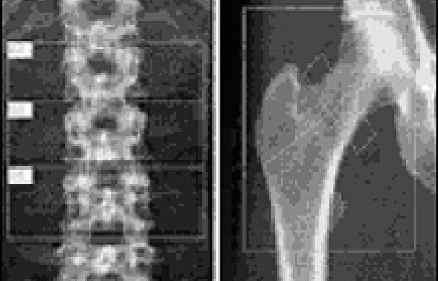
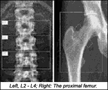
The problem is that as with all modalities, including radiology, DXA is operator-dependent. I don't want to use too many statistics or principles of physics in this article, but there are certain simple facts worth knowing. First, DXA uses X-rays at two energy levels to determine the bone mineral content. This is done by subtracting the difference of the absorption of X-rays between soft tissue and calcified bone. The software program calculates the bone mineral density (BMD) by dividing the bone mineral content by the area of the region of interest (e.g., an area in the spine or neck of the femur). The BMD measured by the program is compared to reference data specific to the scanner that determines the T- and Z- scores.
The World Health Organization (WHO) lists four diagnostic categories on the basis of the T-score that compares the patient's BMD with the mean value in young white women, and is reported in standard deviations above or below the mean. Remember your statistics! Male and ethnic values are available, but still limited. The Z-score compares the patient's BMD with the mean value in a population of similar age, sex and height. The databases for these measurements are not so reliable, because initially, these data or criteria were based on studies in elderly white women, which does not address the rest of the population, (i.e., males, nonwhites, and children). Please keep this in mind when you review a DXA report on a patient other than an elderly white female.
Other risk factors for T -1.5 patients include estrogen deficiency, calcium deficiency, vitamin D deficiency, low body weight, alcoholism, medication use (especially steroids), and smoking. Other risk factors that are not modifiable are female gender, advancing age, white or Asian ethnicity, a family history of osteoporosis, previous fractures and frail health. Maybe we should include fast-food diets!
World Health Organization (WHO) Criteria for Defining Bone Density:1
Condition
Description
Normal
BMD value within 1 SD of the young adult reference mean (T >= -1.0).
Ostepenia
BMD value of more than 1 SD below the young adult mean, but less than 2.5 SD below this value (-1.0 >T >-2.5).
Osteoporosis
BMD value of 2.5 SD or more below the adult mean value (T <= -2.5).
Established osteoporosis
BMD value of 2.5 SD ormore below the adult mean value (T <= -2.5) in the presence of one or more fragility fractures.
(BMD = bone mineral density; SD = standard deviation.)
Thus far, we have only discussed the T- and Z- scores; but what are the other problems? One is that the positioning for DXA in the hip and spine are of vital importance. Errors can be made in the placement and sizing of the regions of interest, affecting the accuracy of the data. If the region of interest is too large, for example, the BMD will be calculated too low - or the opposite. (BMD is calculated by dividing the bone mineral content by the area measured.) Overlying material in the region of interest, such as degenerative osteophytes, can create added density, resulting in inaccurate bone density measurements.
One more issue to consider is the testing site to which you refer your patients. Is it certified by the International Society for Clinical Densitometry? Does the site have a quality-assurance program and proof that the DXA measurements are reproducible? Do the physician and staff know the precision ratings of the scanner and the technicians? Also important - is the cost for the examination reasonable? You can request only a BMD report. If you don't, the patient may also incur an additional consultation fee from the clinical specialist reviewing the BMD report (which you may or may not need, depending on the situation).
What I hope you digest from this simple article is that we need to consider osteoporosis as a preventable problem. It is recommended that all women 65 years of age and older have at least a DXA to rule out possible osteoporosis; it is also recommended for all white postmenopausal women younger than age 65 who have at least one risk factor in addition to menopause. Men can also develop osteoporosis, as smoking is a major cause of declining bone density in both men and women. Osteoporosis in children is difficult to assess, because normal values for children are still limited. They have different growth rates, but in general, density should increase to a peak mass at somewhere between 20 and 30 years of age. I suggest consulting a specialist regarding children with osteoporosis.
References
Information from assessment of osteoporotic fracture risk and its application to screening for postmenopausal osteoporosis. Report of a WHO Study Group. World Health Organ Tech Rep Ser 1994;843:1-129.
National Osteoporosis Foundation. Physician's guide to prevention and treatment of osteoporosis. Washington, D.C., 1998.
Many relevant diagnostic signs are not performed deliberately by the examiner or by the patient at the examiner’s direction. They are observed as the patient reacts to their condition. Fortin’s finger sign, Minor’s sign, and Vanzetti’s sign are three examples of this principle.
Spearheaded by burgeoning scientific and clinical research literature, psychedelics have reached a level of media coverage and popular interest that has not been seen for over half a century. By “psychedelics,” we are referring to the unique class of substances that includes psilocybin (the active compound found in so-called “magic mushrooms”), LSD, dimethyltryptamine (DMT), ayahuasca, 5-MeO-DMT, and mescaline – each of which occurs in the natural world (except for LSD, which is a semi-synthetic compound).
While working at the Lovelace Medical Center in Albuquerque, N.M., one of the primary care physicians stated that imaging every patient prior to receiving chiropractic was not medically necessary. It was at that point I reconsidered the need for the imaging of every patient to avoid malpractice.