What's Your Diagnosis? Pain After Total Hip Arthroplasty
Deborah Pate, DC, DACBR
A patient presents with complaints of hip pain primarily on the right side, lower back pain and stiffness in both hips. She has not been to see the orthopedist who did the surgery on her right hip since the initial surgery and rehab. She states she has not fallen, and that she is able to walk, sit and climb stairs, since she had both hips replaced.
The patient has received chiropractic care in the past, but reports she was advised not to go to chiropractors since she had her hip replacements.
Radiographic Findings
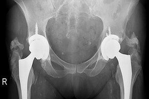
Figure 1 is an X-ray taken to evaluate the status of the hip replacements. The film demonstrates heterotopic ossification involving both hips. There is atherosclerotic calcific plaquing in the femoral arteries, and other calcifications in the pelvic basin including phleboliths, calcific plaquing in the iliac arteries, mesenteric lymph node calcification and/or uterine fibroid.
There is a problem with this AP view. The end of the stem of the femoral component of the THA is not visualized on either side. What should be done is either take an AP hip of each hip to assess the position of the stem or take the same view, but position the focal spot lower down to include the stems. The distal femoral component needs to be visualized to rule out possible loosening.
FIG 1 78 y/o female with right hip total hip arthroplasty two years prior (left THA done four years before the right).
However, should we even consider treating this patient? Before you answer, let's discuss heterotopic ossification (HO), a known complication of total hip arthroplasty. Prevalence can vary widely from 15-90 percent. Male patients with a previous history of HO are said to be at the highest risk.
Diagnostic Criteria
Diagnosis is based on a single AP radiograph and is most commonly done using the Brooker classification, which categorizes HO into four grades, of which grades III and IV are considered clinically relevant. (Grades I and II often are not associated with any major clinical symptoms.)1
Grade I: Described as islands of bone within the soft tissues about the hip.
Grade II: Consisting of bone spurs originating from the pelvis or proximal end of the femur, leaving at least 1 cm between opposing bone surfaces.
Grade III: Consisting of bone spurs originating from the pelvis or proximal end of the femur, reducing the space between opposing bone surfaces to less than 1 cm.
Grade IV: Hip joint ankylosis.
Confirmation & Treatment
The confirmative test is the bone scan. Many bone metabolic turnover markers have been tested, but none has been found to be reliable to date, either with preventing or diagnosing HO. The most effective prophylactic treatment is radiotherapy or administration of nonsteroidal anti-inflammatory drugs. The most commonly prescribed drug in prophylaxis of HO is indomethacin.2 The prophylactic radiation treatment is done preoperatively within 24 hours or postoperatively within 72 hours.3
These are prophylactic treatments; what is the treatment for patients who already have HO causing significant symptoms? Unfortunately, the one and only treatment for HO once it has developed and is at a grade III or IV – associated with significant restriction of motion or ankyloses of the joint, rendering the arthroplasty useless – is a revision arthroplasty.4
Potential Causative Factors
The etiopathogenesis of HO is not completely understood. The development of HO following hip replacement surgery is affected by many factors; some relate to the patient's own risk factors, as well as external factors such as surgical approach, type of implant and length of surgery.
HO is usually evident from radiographs by six weeks after surgery. Hence, the ossification matures throughout the first six months and then generally does not develop further.
Chiropractic Intervention
How many of your patients have hip replacements? I think it is important for us to be aware of this complication when we treat patients with total hip replacements. We need to document our treatment, which means knowing how to diagnose and report the findings.
Allopathic medicine has little to offer patients with grades I or II OH. This is an important issue. Even the when a grade III or IV is present, if the patient still has enough mobility, revision is discouraged because the surgery itself is a factor in the development of OH.
Chiropractic has much to offer these patients: pain relief, better function and range of motion, for starters. We need to document our treatment in the literature. (Here is an example of a case report: "Chiropractic Management of Hip Pain After Conservative Hip Arthroplasty," by Jeffrey Wisdo, DC: https://www.jmptonline.org/article/S0161-4754(04)00133-2/fulltext. Thank you, Dr. Wisdo!)
Back to Our Patient
Are we going to treat this 78-year-old patient with bilateral THA? Will she gain any benefit from our treatment? What grade does she present with per the Brooker classification? I'm confident about grading the right hip, but not quite certain about the left. I would need to evaluate clinical findings, including range of motion; and obtain images of the total hip prosthesis to rule out mechanical loosening. A bone scan would determine if heterotopic bone growth is still active.
I believe chiropractic has something to offer this patient, while her options are limited in regard to allopathic treatment. I would not want to be the one determining whether the left hip needs a surgical revision. That should be left to the orthopedic surgeon and the patient. I would certainly refer this patient to her orthopedic surgeon for an evaluation.
Most surgeons would welcome an option of conservative care before surgical intervention, particularly when it involves a revision. Whenever possible, a team approach is the best option.
Clinical Takeaway
Total hip arthroplasty (THA) has been described as "the operation of the 20th century" for its high satisfaction rates and improvement in quality of life following surgery.5 More than 1 million THAs are performed worldwide per year. Clinical outcome and implant functioning are excellent over time.6
The number of implants is projected to increase by 174 percent in the United States by 2030, and much higher in emerging countries, with worldwide prevalence projected to double within the next two decades.
We have patients with total joint replacements. We are managing their rehabilitation and helping them regain their joint function. This needs to be documented and reported in the literature. Cooperation and collaboration with the best interest of the patient in mind is where our health care system needs to be.
References
Cai L, Wang Z, Luo 1, et al. Optimal strategies for the prevention of heterotopic ossification after total hip arthroplasty: a network meta-analysis. Int J Surg, 2019 Feb;62:74-85.
Lee A, Amin NP. Radiation therapy for heterotopic ossification prophylaxis. In: StatPearls [Internet]. Treasure Island (FL): StatPearls Publishing; 2019.
Å?egosz P, Otworowski M, Aleksandra S, et al. Heterotopic ossification: a challenging complication of total hip arthroplasty: risk factors, diagnosis, prophylaxis, and treatment. BioMed Res Intern, 2019.
Biz C, Pavan D, Frizziero A, Baban A, Iacobellis C. Heterotopic ossification following hip arthroplasty: a comparative radiographic study about its development with the use of three different kinds of implants. J Orthop Surg Res, 2015;10:176.
Zagra L. Advances in hip arthroplasty surgery: what is justified? EFORT Open Rev, 2017;2(5):171–178.
Hip and Knee Arthroplasty: Annual Report 2015. Australian Orthopaedic Association National Joint Replacement Registry.
Many relevant diagnostic signs are not performed deliberately by the examiner or by the patient at the examiner’s direction. They are observed as the patient reacts to their condition. Fortin’s finger sign, Minor’s sign, and Vanzetti’s sign are three examples of this principle.
Spearheaded by burgeoning scientific and clinical research literature, psychedelics have reached a level of media coverage and popular interest that has not been seen for over half a century. By “psychedelics,” we are referring to the unique class of substances that includes psilocybin (the active compound found in so-called “magic mushrooms”), LSD, dimethyltryptamine (DMT), ayahuasca, 5-MeO-DMT, and mescaline – each of which occurs in the natural world (except for LSD, which is a semi-synthetic compound).
While working at the Lovelace Medical Center in Albuquerque, N.M., one of the primary care physicians stated that imaging every patient prior to receiving chiropractic was not medically necessary. It was at that point I reconsidered the need for the imaging of every patient to avoid malpractice.