Conservative Management of Hamstring Strains, Part 1
Thomas Michaud, DC
Of all the gait-related muscle injuries, hamstring strains have the highest rate of recurrence, with as many as one-third of injured athletes suffering reinjury within the first few weeks following return to sport.1 Because their stride lengths may exceed 3.5 meters, sprinters are especially vulnerable to this injury, and it can take up to four months of aggressive rehabilitative exercises before return to sport is possible.2
Hamstring Anatomy
The hamstrings are comprised of four separate muscles: semitendinosus, semimembranosus, and the long and short heads of the biceps femoris muscle. The semitendinosus and semimembranosus originate from the medial aspect of the ischial tuberosity, while the long head of the biceps femoris originates from the lateral ischial tuberosity and the sacrotuberous ligament. (Fig. 1)
The short head of the biceps femoris originates from the lateral intermuscular septum and the distal one-third of the lateral femoral cortex. Distally, the semitendinosus attaches to the medial tibia on the lower portion of the pes anserine muscle group, where it is thought to play a role in assisting gastrocnemius with knee flexion during propulsion.
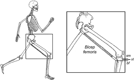
Fig. 2: Because the distal biceps femoris has a lower point of attachment than the semitendinosus (st) and semimembranosus (sm), forward motion of the leg during swing phase creates a greater tensile strain in the biceps femoris muscle. The increased tension allows the biceps femoris to stabilize the sacroiliac joint during early stance phase. Modified and redrawn from Thelen, et al.14
The semimembranosus is a much more complicated muscle, as it possesses five separate attachments that allow it to play an important role in decelerating anterior tibial translation and valgus collapse of the knee during stance phase. Because it has a direct attachment to the posterior horn of the medial meniscus, the semimembranosus muscle protects the meniscus by displacing it posteriorly upon knee flexion.
While the semimembranosus and semitendinosus are important stance-phase stabilizers, the long and short heads of the biceps femoris muscle, owing to their more distal attachment on the proximal fibula, are important decelerators of forward motion of the swing-phase leg. (Fig. 2) The long head of the biceps femoris also plays an important role in stabilizing the sacroiliac joint, and in lessening bony / soft-tissue vibrations during contact.
Injury Mechanisms
Given their dissimilar functions, the various hamstring muscles are injured through different biomechanical mechanisms. The semimembranosus is more likely to be strained with activities that stretch the hamstrings through extreme ranges of motion, with the classic mechanism being a full front or side split. This injury is more likely to occur in sports like ballet, gymnastics, cheerleading and even yoga, during which a slow forward or side split may overstretch and tear the medial hamstrings. Stretch injury can also occur with a fast motion, such as the rapid high kicks associated with various martial arts.
As demonstrated by Askling, et al.,3 stretch injuries frequently injure multiple muscles at the same time. In fact, in their detailed MRI study of 30 stretch-related hamstring injuries in athletes from 21 different sports, Askling, et al., noted that the semimembranosus muscle was injured in 83 percent of subjects, while the quadratus femoris, semitendinosus, biceps femoris long head, and adductor magnus muscles were also injured, but at a much lower frequency.
The authors correlate the severity of hamstring injury and subsequent rehabilitation time directly to the proximity of injury to the ischial tuberosity: the more proximal the injury, the more difficult it is to treat. The severity of these injuries is underscored by the fact that 47 percent of the injured athletes were unable to resume their sports career.
Unlike the more extensive stretch-related medial hamstring injuries, runners tend to strain primarily the long head of the biceps femoris muscle. Because it possesses a longer lever arm for decelerating knee extension, this muscle is usually injured just prior to heel strike, when excessive tensile strain present in the long head results in failure of the proximal muscle tendon junction. The biceps femoris muscle is typically injured when switching from the eccentric contraction present during midswing, to the nearly isometric contraction present during late swing, when the tendons of the long head of the biceps femoris muscle are stretching in order to store and return energy.
Since running-related biceps femoris injuries tend to occur at the proximal muscle tendon junction, the recovery time for this injury is faster than the stretch-related semimembranosus injuries, which are more likely to damage the tendon itself. A biceps femoris injury can be identified by palpating the muscle-tendon junction slightly distal to the lateral ischial tuberosity, and by having the patient gently isometrically tense the muscle.
Risk Factors
There are several modifiable and nonmodifiable risk factors responsible for biceps femoris injuries. As listed by Brooks, et al.,4 nonmodifiable risk factors include previous hamstring injury, older age, and black or aboriginal ethnicity; while modifiable risk factors include quadriceps / hamstring strength imbalance, inadequate warm-up, muscle stiffness, poor lumbopelvic strength / stability, and the presence of a hyperlordotic lumbar spine with an anteriorly tilted pelvis. The latter postural factors may result in hamstring injury because a tilted pelvis shifts the ischial tuberosity superiorly, increasing tensile strain in the hamstring origin.
As with most injuries, the single best predictor of future injury is prior injury, which may increase muscle stiffness and alter neuromotor control. Because of the extremely high rate of recurrence associated with biceps femoris strains, rehabilitation of this injury must be comprehensive and address all possible factors associated with chronicity.
While hamstring inflexibility is often cited as a potential cause, there is little evidence to support this theory.5 In fact, excessive stretching is more likely to cause a hamstring injury than alleviate one, particularly during the acute phase following injury. In Askling, et al.'s detailed study of hamstring injuries in different types of athletes, the only running-related hamstring strain reported occurred while the athlete was stretching before participating in sport. Subsequent MRI confirmed the runner tore both the semimembranosus and biceps femoris long head muscles.
References
Orchard J, Best T. The management of muscle strain injuries: an early return versus the risk of recurrence. Clin J Sport Med, 2002;12:3-5.
Askling C, Saartok T, Thorstensson A. Type of acute hamstring strain affects flexibility, strength, and time to return to pre-injury level. Br J Sports Med, 2006;40:40.
Askling C, Tengvar M, Saartok T, Thorstensson. Proximal hamstring strains of stretching type in different sports. Injury situations, clinical and magnetic resonance imaging characteristics, and return to sport. Am J Sports Med, 2008;36:1799.
Brooks J, Fuller C, Kemp S, et al. Incidence, risk, and prevention of hamstring muscle injuries in professional rugby union. Am J Sports Med, 2006;34:1297.
McHugh M, Connolly D, Eston R, et al. The role of passive muscle stiffness and symptoms of exercise-induced muscle damage. Am J Sports Med, 1999;27:594.
Part 2 of this article discusses treatment and prevention of hamstring injuries; it is scheduled to run in the June 17 issue.
Many relevant diagnostic signs are not performed deliberately by the examiner or by the patient at the examiner’s direction. They are observed as the patient reacts to their condition. Fortin’s finger sign, Minor’s sign, and Vanzetti’s sign are three examples of this principle.
Spearheaded by burgeoning scientific and clinical research literature, psychedelics have reached a level of media coverage and popular interest that has not been seen for over half a century. By “psychedelics,” we are referring to the unique class of substances that includes psilocybin (the active compound found in so-called “magic mushrooms”), LSD, dimethyltryptamine (DMT), ayahuasca, 5-MeO-DMT, and mescaline – each of which occurs in the natural world (except for LSD, which is a semi-synthetic compound).
While working at the Lovelace Medical Center in Albuquerque, N.M., one of the primary care physicians stated that imaging every patient prior to receiving chiropractic was not medically necessary. It was at that point I reconsidered the need for the imaging of every patient to avoid malpractice.