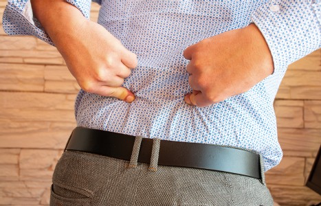
Many relevant diagnostic signs are not performed deliberately by the examiner or by the patient at the examiner’s direction. They are observed as the patient reacts to their condition. Fortin’s finger sign, Minor’s sign, and Vanzetti’s sign are three examples of this principle.
Latest Information on Why Friction Massage Heals Tendinitis
In manual therapy, clinical results usually precede the scientific validation. Most techniques are used because "it works." Usually the author of the method hypothesizes why it works, but the reasoning although appearing logical may have nothing to do with the result. According to Frank and Hart1: "Mechanical loading influences cell behavior in all soft tissues, but how it does so is not fully understood." The latest research on the effect of loading or deep pressure has been directed to the cellular level. In 1979, Meikle et al.2 found that continuous stress to newborn rabbit cranial sutures led to an increase in collagen synthesis. Fibroblasts are responsible for increased collagen synthesis. Slack et al.,3 using a model of cyclic tensile loading of isolated embryonic chick tendons in vitro, showed increased synthesis of proteins, glycosaminoglycans and DNA by the fibroblasts.
Healing of tendons occurs in three stages. In the inflammatory stage, platelets and fibrin fill the wound and fibroblasts and phagocytic cells migrate to the injured area. Fibroblasts produce fibronectin, which acts as an adhesive molecule to bind collagen. As healing proceeds the fibroblastic production of fibronectin decreases. In the proliferative stage, the fibroblasts increase in number and synthesize collagen. Finally, in the third stage of remodeling or the maturation stage. there is a realignment of collagen fibers, and the collagen production shifts from the immature Type III to the mature Type I collagen.
Davidson et al.4 recently (1997) created a tendinitis in the rat tendon by injecting the enzyme collagenase. This method has also been used to trigger an inflammatory response in horses and ponies. While the collagenase did not cause the typical inflammatory response with the appearance of numerous mononuclear blood cells and lymphocytes at the injured site, the hallmark of tendon injury which is collagen fiber disruption and misalignment was exhibited in the rat tendons. They used augmented soft tissue mobilization (ASTM), which is an aluminum instrument used to apply "considerable pressure" to a tendon without breaking the overlying skin. The tendon was massaged longitudinally moving distal to proximal and proximal to distal along the length of the Achilles tendons of the rats.
There were four groups of rats: a) control; b) collagenase induced tendinitis; c) collagenase induced tendinitis plus ASTM; and d) ASTM alone to a normal tendon. After injecting the collagenase, the tendons were allowed to heal for three weeks. After the three weeks, ASTM was performed on group C and D for three minutes on postoperative days 21, 25, 29 and 33, i.e., four treatments. One week after the last treatment, the tendons were harvested and evaluated under light and electron microscopy and immunostaining for type I and type III collagen and fibronectin.
Light microscopy showed increased activated fibroblast proliferation in the tendons of group C and D. ASTM proved to initiate fibroblast activation which eventually leads to collagen synthesis. Group C also showed more fibronectin antibodies than group A. There was little fibronectin in Group B, but an increased amount in Group D. Gait analysis was also performed, and a significant improvement in stride length and stride frequency only occurred for Group C between post surgery day 21 and the final observation day.
ASTM promoted healing and earlier recovery of limb function following collagenase injury by the increased fibroblastic proliferation. The earlier recovery allowed increased limb function which also helped in the promotion of fiber realignment.
Of course, more studies are necessary especially to determine the effect of friction on chronic long-term scar tissue. It definitely appears that applying friction massage with increased pressure creates fibroblastic proliferation.
References
- Frank CB, Hart DA. Cellular response to loading. In: Leadbetter WB, Buckwalter JA, Gordon SL. Sports-Induced Inflammation, Amer Acad of Orthop Surgeons, Park Ridge, IL, 1989:555-563.
- Meikle MC, Reynolds JJ, Sellers A, et al. Rabbit cranial sutures in vitro: A new experimental model for studying the response of fibrous joints to mechanical stress. Calcif Tissue Int 1979;28:137-144.
- Slack C, Flint MH, Thompson BM. The effect of tensional load on isolated embryonic chick tendons in organ culture. Connect Tissue Res 1984;12:229-247.
- Davidson CJ, Ganion LR, Gehlsen GM et al. Rat tendon morphologic and functional changes resulting from soft tissue mobilization. Medicine & Science in Sports & Exercise. American College of Sports Md, 1997:313-319.
Warren I. Hammer, DC
Norwalk, Connecticut


