Many relevant diagnostic signs are not performed deliberately by the examiner or by the patient at the examiner’s direction. They are observed as the patient reacts to their condition. Fortin’s finger sign, Minor’s sign, and Vanzetti’s sign are three examples of this principle.
Radiation Exposure From CT Scans: Is There Increasing Cause for Concern?
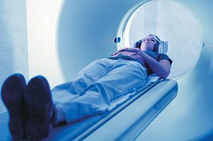
Radiation exposure from medical imaging has become a concern with the dramatic increase in scanning (particularly CT scanning) over the past several decades. In 1980, 3 million CT scans were performed in the U.S.; this has increased to more than 62 million CT scans per year, 4 million of which are performed on children.1 It is estimated that one-third of these scans may not be medically necessary. For example, some emergency departments have drastically increased the use of CT scans for the examination of patients with abdominal pain or headaches.
Another concern is the increase in whole-body CT scans ordered by patients themselves. These scans are marketed to the general public as screening tests and are sometimes routinely repeated. To date, the positive or negative predictive value of these whole-body scans for disease detection has not been determined. And then there is the problem of self-referral; practitioners who own imaging facilities have a tendency to order studies more frequently.1 Medical ionizing radiation has great benefits and should not be feared; however, judicious use of this modality should be followed.
Radiation Doses and Risks
The International Commission on Radiological Protection (ICRP) estimates that the average person has an approximately 4 percent to 5 percent increased relative risk of fatal cancer after receiving a whole-body dose of 1 Sv. Other studies, however, have not agreed with this estimate, estimating it to be much lower, around 1 percent.2 The question of how much radiation is safe has always been difficult to answer. In an effort to quantify these risks, David J. Brenner, PhD, DSc, and Eric J. Hall, DPhil, DSc, of Columbia University, cite studies indicating that 0.4 percent of all cancers in the U.S. might be attributed to radiation exposure.

Extrapolating those results to current CT use, Brenner and Hall believe the current proportion of cancers attributable to CT-associated radiation could be as great as 1.5 percent to 2 percent. Of particular concern is the increased use of CT in pediatric diagnosis, which has exposed children to adult radiation protocols that only recently were modified for pediatric applications.
The cancer risk posed by CT-associated radiation is not fully appreciated by health care professionals or the public. Brenner, and Hall point out that part of the problem is that physicians often view CT studies in the same light as other radiologic procedures, even though CT involves larger radiation doses than conventional X-ray imaging. For example, a conventional abdominal X-ray delivers a radiation dose of 0.25 mSv to the stomach; an abdominal CT exposes the stomach to a dose of 10 mSv. That dose increases to 20 mSv with neonatal abdominal CT.
Estimating cancer risks associated with diagnostic X-rays using epidemiologic tools is difficult because of extrapolation to low radiation doses, recall bias, and different X-ray energies used at various institutions. Most low-dose human ionizing radiation risk estimates come from the atomic bomb survivors in Japan; other supporting studies include a recent large-scale study of 400,000 radiation workers in the nuclear industry - who were exposed to an average dose of approximately 20 mSv.
A significant association was reported between the radiation dose and mortality from cancer in this cohort (with a significant increase in the risk of cancer among workers who received doses between 5 mSv and 150 mSv); the risks were quantitatively consistent with those reported for atomic-bomb survivors. If this evidence is a concern for adults, it should be even more of a concern for children. Children are at a greater risk, both because they are inherently more radiosensitive and because they have more remaining years of life during which a radiation-induced cancer could develop.
Quantifying Cumulative Exposure
Despite the fact that most diagnostic CT scans are associated with very favorable risk-to-benefit ratios, there is a strong case to be made that too many CT studies are being performed in the United States. We need better means of measuring patient exposure and better awareness by both health care practitioners and the general public of the risk of ionizing radiation. Thankfully, the medical community is finally beginning to consider the overuse of diagnostic radiation an important issue. Recently, a new method of tracking radiation doses delivered by CT scans was developed and reported on at the 2010 annual meeting of the American Roentgen Ray Society. George Shih, MD, assistant professor of radiology at Weill Cornell Medical College in New York City, reported on this new system, which extracts radiation dose information from conventional CT scans and puts it into a more user-friendly format. This allows clinicians to determine the cumulative amount of radiation administered to their patients over time. The purpose of the system, called Valkyrie, is to put the information into digital form and "to eventually perform automated quality control, promote radiation safety awareness, and provide a longitudinal record of patient healthcare-related radiation exposure."5
Valkyrie, which Dr. Shih and his colleagues developed in collaboration with radiation physicists at Columbia University, extracts dose information embedded in the images using distinct characteristics that can be reliably detected by established algorithms. No proprietary software is used. Dr. Shih and co-authors tested the system by having it analyze radiation dose information from 518 randomly selected dose reports. Valkyrie processed them all accurately. Although the system is still in the developmental stage, it is now used around the clock at Cornell and is apparently still 100 percent accurate.
This is a very commendable start at monitoring radiation exposure. It remains to be seen if cumulative radiation information will be considered when diagnostic studies are requested. Hopefully the health care community as a whole will take a more active role in reducing patient radiation exposure.
References
- Holmes EB, White Jr. GL, Gaffney DK. "Ionizing Radiation Exposure, Medical Imaging." EMedicine (WebMD), May 9, 2010.
- Brenner DJ, Hall EJ. Computed tomography - an increasing source of radiation exposure. NEJM, Nov. 29, 2007;22(357):2277-2284
- Cardis E, Vrijheid M, Blettner M, et al. The 15-country collaborative study of cancer risk among radiation workers in the nuclear industry: estimates of radiation-related cancer risks. Radiat Res, 2007;167:396-416.
- Cardis E, Vrijheid M, Blettner M, et al. Risk of cancer after low doses of ionising radiation: retrospective cohort study in 15 countries. BMJ, 2005;331:77-77.
- "Valkyrie Computer Program Tracks Cumulative CT-Related Radiation Exposure." Medscape Medical News, May 6, 2010.


