The Shoulder Girdle's Importance to the Cervical Spine
Kevin G. Hearon, DC, CCEP, CCSP
The shoulder girdle consists of three bones: the clavicle, scapula and humerus. It is suspended at its posterior margins from the cervical and thoracic spine by the trapezius muscles, levator scapula, rhomboid, serratus and latissimus dorsi. The anterior muscle attachments include all of the pectoral muscles, subclavius and platysma. While the rib cage offers an attachment site for some of these muscles, it also acts as an articulation over which the scapula may glide in many directions, making it a very mobile and adaptive structure.
Central to the stability of the shoulder girdle is the clavicle, which is the only bone directly attached by joints to the axial skeleton at the sternoclavicular and acromioclavicular locations. This important bone is primarily responsible for keeping the shoulder girdle at proper distance from the thorax, facilitating function and mobility of the upper extremity. When the clavicle gets separated at either end, it compromises the position and function of the whole shoulder girdle complex.
The S/C (sternoclavicular) joint is readily viewed by physical exam or on A-P thoracic X-ray. A 5 mm or greater difference in height signifies an automatic separation of this joint. The mechanoreceptors at this joint will inhibit its adjacent muscular supply. The muscle most easily isolated for testing is the pectoralis major clavicular.
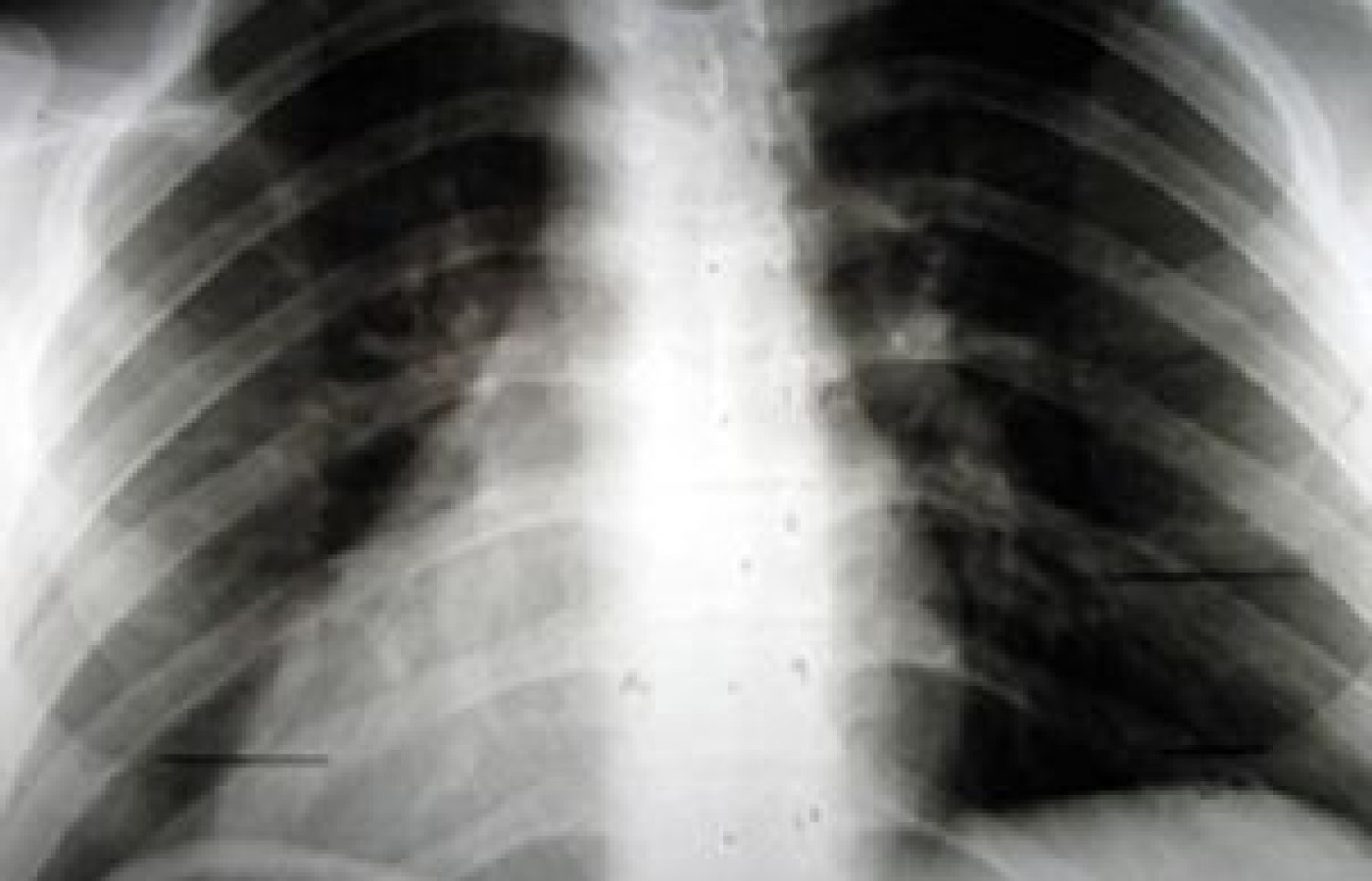
A-P thoracic film demonstrating left cervicobrachial traction syndrome; left inferior S/C of 10 mm, left inferior scapula of 34 mm.
This A-P thoracic film demonstrates a left inferior S/C of 10 mm and a left inferior scapula of 34 mm following a motor-vehicle accident. Diagnostically this is called a left cervicobrachial traction syndrome. This sets the patient up for a constant tug-down on the left side of the cervical spine by the upper trapezius muscle, which would result in chronic neck subluxations.
Correction of this malady is accomplished by restoring the left inferior S/C back to its normal position with an adjustment. Once the adjustment is made, it is then appropriate to post-check the pectoralis major clavicular muscle, which should now be restored to full strength. On the rare occasion that this joint is still weak upon testing and hypermobile, it would then need to be taped twice a week for six weeks to regain stability.
Your notes should now document the reduction of the S/C upon palpation and if necessary on X-ray. In addition, you should note a significant reduction of the scapular height within normal limits. This reduction will greatly reduce the tugging stress of the shoulder girdle upon the cervical spine.
S/C reduction to 2 mm and scapula to 10 mm after adjusting the left inferior S/C back to its normal position.
The S/C is reduced to 2 mm and the scapula to 10 mm by the next day, as proven with X-ray, and the pectoralis major clavicular now tests strong.
The A/C (acromioclavicular) joint is the distal end of the clavicle and has a reputation of being frequently unstable. Grading the level of sprain for this joint, in my opinion, is of higher value than the S/C due to its instability tendency. Normal coracoid to clavicle distance is 1.1 to 1.3 cm. Anything above 1.3 cm is up for consideration of an A/C separation. When watching sports on TV, it is not uncommon to hear description of an athlete's injury of the shoulder as either a dislocation, which is glenohumeral, or a separation, which is an A/C joint.
Due to the scapula dropping away, it is once again important to check for a scapula height difference of 15 mm or greater, which would be considered a shoulder girdle instability.
The muscle most easily isolated for testing this joint that is inhibited when subluxated is the coracobrachialis muscle in the first 30 degrees of forward flexion. It is better to test it in the supine position due to its strength.
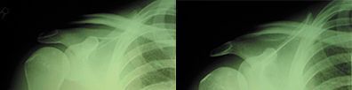
One of the myths out there that is still being taught is that when an X-ray is taken of an A/C separation, the patient should hold a 5 lb. weight in their hand, and this will enhance the separation on the film. This could not be further from the truth. Here are weighted and unweighted films of an A/C separation; you can decide for yourself.
This patient would obviously be a grade 3 separation with a significant horizon sign. The coracobrachialis muscle would be very weak and the shoulder is significantly drooping on this side.
Lateral compression into the shoulder girdle is the No. 1 impact of injury creating this condition. Numerous sports are loaded with this type of impact; therefore, it is a very common injury.
L-R: Unweighted and weighted films demonstrating an A/C separation.
Horizon sign is a term used to describe the lump along the top surface of the shoulder A/C that is more prominent than the normal side. A grade 1 A/C subluxation has no horizon sign and is weak upon testing the coracobrachialis. Grade 2 A/C separation has a horizon sign and the clavicle will move down in relation to the acromion while performing the coracobrachialis test. A grade 3 A/C separation has a horizon sign and will not move in relation to the acromion while performing the coracobrachialis test.
Adjusting an A/C separation is a fairly gentle procedure from superior to inferior. A grade 1 subluxation does not need stabilization. Grade 2 and grade 3 typically need to be either taped or supported with a Sully A/C stabilizer for a period of six weeks to become stable. Even old A/C separations of many years' duration frequently respond beautifully to this type of care.
Please remember that this joint will create unequal pulling on the cervical spine due to scapula height variation and result in chronic cervical subluxations unless it is taken care of. So, when it comes to creating a good environment for stability of the cervical spine it would be wise to consider the shoulder girdle as a possible contributing factor in its treatment.
Many relevant diagnostic signs are not performed deliberately by the examiner or by the patient at the examiner’s direction. They are observed as the patient reacts to their condition. Fortin’s finger sign, Minor’s sign, and Vanzetti’s sign are three examples of this principle.
Spearheaded by burgeoning scientific and clinical research literature, psychedelics have reached a level of media coverage and popular interest that has not been seen for over half a century. By “psychedelics,” we are referring to the unique class of substances that includes psilocybin (the active compound found in so-called “magic mushrooms”), LSD, dimethyltryptamine (DMT), ayahuasca, 5-MeO-DMT, and mescaline – each of which occurs in the natural world (except for LSD, which is a semi-synthetic compound).
While working at the Lovelace Medical Center in Albuquerque, N.M., one of the primary care physicians stated that imaging every patient prior to receiving chiropractic was not medically necessary. It was at that point I reconsidered the need for the imaging of every patient to avoid malpractice.