Biomechanics: The Swiss Army Knife of Forensic Evaluation
Arthur Croft, DC, MS, MPH, FACO
One of the most frustrating aspects of patient care is watching your patient's legal claim go down in flames. Often they become saddled with your medical bill and others they expected the responsible insurer would pay. Adding insult to injury, many states have a statutory offer system in place whereby one party must pay the other party's legal fees if they fail to recover more than what was offered to them prior to an arbitration or trial. This is just one of the factors driving plaintiffs to accept low settlement offers. The fallout often affects the treating doctors, who are then asked to take a cut in their fees.
Sound familiar? It's a common outcome of motor vehicle collision (MVC) injuries involving third-party payors. But the solution is definitely not to walk away. Far from it. These patients desperately need your skilled hands, for one thing, and personal injury should now be one of the more lucrative aspects of your practice.
It's time to make a case for shoring up the troubled personal-injury arena in ways DCs are particularly skilled in: biomechanics. In addition to biomechanics knowledge, of course, you can also provide a risk analysis and what I call these days biomedical testimony: discussing injuries, diagnoses, appropriateness of care, MRI findings, prognosis, etc. And consider this: key in the intimidation arsenal of the defense attorney are the experts they wield - accident reconstructionists, biomechanists, and various medical or chiropractic examiners.
To counter them, the plaintiff attorney much hire a matching panel of experts at a formidable cost. If not, they can expect to be seriously outgunned at trial. If these experts do come on board, but the case goes sour anyway, the plaintiff could be faced with even steeper bills. So, by wearing several experts' hats, you become a very attractive treater/expert indeed. And your fees for forensic work will never be subject to Medicare manipulations or any other reimbursement changes.
Why is biomechanics so critical? Because a critical point of attack by the defense in many cases is causation. Your patient may have been in fairly good health before the crash, but then, failing conservative care, might have undergone a spinal fusion. The classic defense argument would be, "Hey look, he's a 49-year-old man with pre-existing degenerative changes. He would have needed that surgery anyway, and the MVC was just a coincidence. He's just using the car crash to bankroll his surgery." And more often than not, when the surgeon is asked whether the MVC caused the changes that required surgery, they will have no firm opinion.
Many defense verdicts result from a skillful exploitation of this kind of weak spot. And this is where biomechanical testimony or expertise becomes so important. In the process of connecting the dots, you can analyze the crash, move on to the biomechanical features and probability of injury, and then correlate it with the outcome. Finally, you can do a risk analysis, which is the epidemiological component. That's four experts in one, if you're keeping count. The following is an unusual case that illustrates what can be done with simple biomechanical analysis.
A Case of Chronic Lateral Epicondylitis
A middle-aged man was a belted front seat passenger in a large passenger vehicle traveling at highway speed on a typical southern California freeway. They were hit from the rear by another vehicle, causing the driver to lose control, veer to the left, cross three lanes of traffic, and strike the concrete center divider at a speed the man estimated to be 40 mph. Initially, his chief complaints were chest and neck pain, but not long after the crash he developed right lateral epicondylitis (LE). Over time the chest and neck complaints resolved, but the elbow pain became intractable. An MRI revealed tearing of the extensor carpi radialis brevis muscle (ECRB) at the origin. After three injections of steroids and a variety of unsuccessful conservative approaches, he faced the prospect of surgical exploration and repair.
The defense orthopaedic surgeon's opinion was that the LE was consistent with the man's athletic lifestyle and physical work requirements, and that it could not have resulted from the collision in any case. There were no prior medical records of this complaint. The defense doctor's opinion was, as is often the case, purely speculative.
I was provided repair records for the vehicle and numerous photographs, as well as the MRI, the medical records, and the police report. The first task in this kind of analysis is to establish a reasonable range of crash speeds. The damage profile to the car made it clear that the collision was slightly oblique, but close to a perpendicular one with the center concrete divider. That means that the collision was very comparable to a Federal Motor Vehicle Safety Standard (FMVSS) 208 compliance test. These have, in the past, been conducted at 30 mph into rigid barriers and the data are publicly available. (Now they are conducted at 35 mph.) Based on comparative photographs of similar crash tests, it appeared that the collision would have been somewhat more severe in this case. Without going into the details as to how, the mean delta V (speed change) I arrived at was 32 mph.
I then looked at dummy crash tests conducted at these speeds and in an oblique loading condition. Examination of Figure 1 shows that the chest loads do not begin to significantly ramp up until about 30 msec (30 thousandths of a second). Newton's first law of motion tells us that an object (the plaintiff in this case) traveling at 32 mph, will tend to continue to move at that speed and in the same direction unless acted upon by a net force. In this case, as the car stops after hitting the divider, he continues forward toward the dashboard. Because he's wearing his restraints, they will arrest that motion, but there are two factors that retard this response by about 30 msec: (1) slack in the webbing; and (2) time needed to engage the belt locking mechanism. At the top end of the loading, the belt will actually stretch when the tensile load reaches about 7 kN.
Figure 1. Belt loads are not significantly developed in frontal FMVSS 208 compliance tests until about 30 msec after impact when all slack is removed. (Adapted from Babu V, et al., 2003.1)
The passenger is holding the door grip with the hand of the arm that later developed LE and is squeezing it rather frantically in preparation for the impending collision. So, at this point, we can begin to see that for about 30 msec after the crash, a significant amount of force would have been exerted on the elbow joint because his hand contact and the door grip are relatively rigid. Yet the mass of his torso is decelerating forward against that contact. Because the feet are planted against the floor pan, we can assume that he could dissipate some of this load by bracing there. So, let's find out what these forces were. This can be easily done using the impulse-momentum theorem as follows.
Momentum, p, is equal to mass, m, times velocity, v. We have here a 210 lb (95 kg) man and a velocity of 32 mph (14.3 m/s). And let's subtract his pelvis and lower extremities (about one-third of his body weight) based on the assumption of bracing with his feet:
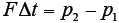
According to the impulse momentum theorem, the change in momentum, delta p, must initially be equal to the impulse exerted by the door hand grip (first equation below); the net force, F, can then be calculated as follows (second equation below):
Since the velocity at p2 is zero, that momentum is also zero. This force is equal to -6,857 lb. To deal with the uncertainty of the numbers in this analysis, I applied a statistical method know as a Monte Carlo simulation. This is a highly processor-intensive method that was actually used to solve the quantum dynamics equations during the development of the first nuclear weapons used in World War II. It's hard to imagine that in those days, they used slide rules. A simpler approach to uncertainty analysis is to simply run this analysis for the most and least conservative crash severities and assume that the true value lies somewhere within that range.
There are other somewhat more rigorous mathematical methods one can use as well, ranging from simple algebraic ones to the more elegant application of differential calculus (e.g., a Taylor series expansion). It can be demonstrated, interestingly, that the more elegant methods are not much more precise in this application.
At this point we have a mechanism for a very high-loading impulse to the joint, and now we should ask ourselves whether this is the kind of load path that is likely to result in microtearing of the ECRB attachment tissue. Perhaps surprisingly, the answer is "probably not." This suggests two things: (1) that we consider an alternative form of physical injury; or (2) that we consider an alternative mechanism of injury.
In the first case, looking again at the MRI, I noticed that there was clearly some joint effusion. This is usually a sign of direct joint injury. That would be a likely consequence of this loading path. Thus, the tearing of the ECRB might not be the primary or solitary source of elbow pain. But what about an alternative load path or injury mechanism
Figure 2. The ECRB muscle and tendinous attachments to the dorsum of the hand. Its chief action is extension of the wrist.
What is it that causes a muscle to tear at its origin? Typically it is a high-force eccentric contraction. Tearing of muscle does not occur during concentric contraction. If it did, muscular strains would be very common, especially in athletes and laborers. Human skeletal muscle is not capable, under normal physiological conditions, of generating a force of sufficient magnitude to cause tearing of fibers. It is a kind of physiological fail-safe. But, when contacting forcefully, if the joint motion is suddenly reversed, this can add sufficient force to rupture muscle fibers. We see this sometimes in sports when sprinters dorsiflex the foot as the calf muscles are violently contracting to produce plantar flexion
Figure 3. Mechanical drawing of the right hand gripping the door handle and then flexing slightly under the load of the forward-moving torso.Figure 4. The principal direction of force (PDOF) is a term commonly applied in accident reconstruction. More correctly, it describes the line of the impulse. This impact will be associated with a counterclockwise rotation of the vehicle and will force the passenger into the door panel.
The tendon of the ECRB attaches to the dorsum of the hand and the tendon is capable of withstanding greater force than the muscle. The muscle is chiefly an extensor of the wrist, as illustrated in Figure 2. In order to place an eccentric load on that muscle, the wrist would have to flex slightly. Could this have happened during this collision? In the plaintiff's case, as he was actively contracting the muscle, he suddenly decelerated against this door grip and the line of action of his torso's inertia would have been down the forearm to the wrist, and would have more than likely forced the wrist into some degree of flexion because the collision was slightly oblique. (Figure 3) This would have produced a forced eccentric load on the strongly contracting muscle and would have resulted in the tearing of the attachment. The crash is drawn schematically in Figure 4.
Finally, I would take into consideration that important material property of all soft and hard tissues: viscoelasticity. Soft tissues are nonlinear, inhomogenous and anisotropic. Their ability to withstand certain forces is rate dependent and means, in plain English, that the stiffness of the tissue is increased under very high loading rates. This will further increase the probability of injury.
Ultimately there were probably two mechanisms of injury at work in this case: one to the joint itself, from compressive loading, and one to the ECRB muscle attaching to the joint as a result of violent eccentric contraction.
At this point we have developed a well-grounded theory for the biomechanical cause-and-effect relationship between the MVC and its outcome through a relatively simple, qualitative reconstruction, and a simple biomechanical analysis, by applying what we know about soft-tissue injuries and how they occur.
References
Babu V, Kevin R, Thomson KR, Sakatis C. LS-DYNA 3D interface component analysis to predict FMVSS 208 occcupant responses. SAE Tech Paper Series, 2003;2003-01-1294.
The proposed merger of the National Board of Chiropractic Examiners and Federation of Chiropractic Licensing Boards was approved by NBCE delegates and FCLB members at their respective annual meetings, held jointly in Atlanta, Ga., this year. Per the new bylaws, the new entity takes the NBCE name, with FCLB continuing as a department within NBCE. The federation will continue to enjoy Board of Directors representation on what will be a single, expanded board.
Before introducing subscapularis syndrome as an upper extremity analog, it is essential to revisit piriformis syndrome as a well-established example of myogenic pseudo-radiculopathy. Piriformis syndrome has long served as a clinical exception to disc-centric models of lower extremity pain and provides an important framework for understanding how deep muscular dysfunction can mimic radiculopathy in the absence of nerve root compression.
Pain has become the dominant language of musculoskeletal healthcare. Numeric pain-rating scales and symptom reports are routinely used as primary indicators of clinical success. But while pain reduction is meaningful, it is an incomplete and often misleading representation of recovery. This has real consequences for patient adherence, long-term outcomes, and how conservative care is perceived within the broader healthcare system.