Accessory ossicles in the foot are abundant, making it difficult to assess whether or not there is a fracture.
os talotibiale
os supratalare
an avulsion, not an accessory bone
os supranaviculare
os infranaviculare
os intercuneiforme
os cuneometatarsale II dorsale (also os cuneometatarsale dorsale fibulare)
os intermetatarsale
os unci? Coalescing with the fourth metatarsal bone?
accessory skeletal element distal to the cuboid (wandering os unci?)
secondary cuboid (cuboides secundarium)
calcaneus secundarius
os tibiale externum
trigonum
os accessorium supracalcaneum
posterior calcaneal spur, not an accessory bone
os subcalcis
inferior calcaneal spur, not an assessory bone
bursitis, not an accessory bone
peroneal bone (os peroneum)
os vesalianum
Most of the time a simple evaluation of the opposite foot will allow one to differentiate most fractures from an accessory ossicle.
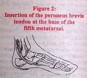
One of the most commonly missed fractures that can be mistaken as an accessory ossicle is an avulsion fracture at the base of the fifth metatarsal. Avulsion of the styloid process of the fifth metatarsal are caused by an inversion injury to the ankle, stressing the peroneus brevis tendon which causes an avulsion at the insertion of this tendon.
Usually the patient will describe a history of stepping off a curb or falling down the stairs. Clinically, this fracture initially appears to be an injury to the ankle, due to the marked swelling which occurs over the lateral aspect of the ankle. If only ankle films are taken, this fracture will be missed.
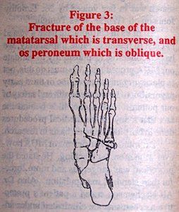
This fracture can also be confused with the os peroneum. Radiographically these two entities can be differentiated by the direction of the fracture line. The fracture line is usually oriented transversely in relationship to the base of the matatarsal. In contrast, when an os peroneum is present, the line of separation is usually obliquely longitudinal in relationship to the base of the metatarsal.
If the fracture is non-displaced, a compression bandage can be used along with restricting weight-bearing until the edema has resolved. If the fracture is displaced, the ankle and foot should be immobilized in the neutral position. Referral to an orthopedist or podiatrist would be recommended for proper management unless the chiropractor is proficient in casting techniques and fracture management. Ice and pulsed ultrasound also can be very helpful in reducing the edema. It should be noted that if the fracture remains open after nine months and is still symptomatic, surgical intervention may be necessary.
Deborah Pate, D.C., DACBR San Diego, California
Editor's Note:
Dr. Pate's book, Case Studies in Chiropractic Radiology is now available through MPI's Preferred Reading and Viewing list. Please see pages xx, Part #T123 for further information on how to order your copy.
The proposed merger of the National Board of Chiropractic Examiners and Federation of Chiropractic Licensing Boards was approved by NBCE delegates and FCLB members at their respective annual meetings, held jointly in Atlanta, Ga., this year. Per the new bylaws, the new entity takes the NBCE name, with FCLB continuing as a department within NBCE. The federation will continue to enjoy Board of Directors representation on what will be a single, expanded board.
Before introducing subscapularis syndrome as an upper extremity analog, it is essential to revisit piriformis syndrome as a well-established example of myogenic pseudo-radiculopathy. Piriformis syndrome has long served as a clinical exception to disc-centric models of lower extremity pain and provides an important framework for understanding how deep muscular dysfunction can mimic radiculopathy in the absence of nerve root compression.
Pain has become the dominant language of musculoskeletal healthcare. Numeric pain-rating scales and symptom reports are routinely used as primary indicators of clinical success. But while pain reduction is meaningful, it is an incomplete and often misleading representation of recovery. This has real consequences for patient adherence, long-term outcomes, and how conservative care is perceived within the broader healthcare system.