When I took a course in drop table methods in the early 1980s as a chiropractic student, we spent most of the course puzzling through the intricacies of the Derifield leg check. The drop table adjustive procedures, by comparison, were a piece of cake. Poring through the Thompson Technique manual, I learned for the first time what it meant to use a chiropractic technique system.
The Derifield Leg Check System
Cervical syndrome: turning the head in the prone position balances leg lengths that appeared uneven, or unbalances leg lengths that appeared even
Pelvic negative syndrome (D-): a knees-extended apparent short leg remains short when the knees are flexed
Pelvic positive syndrome (D+): a knees-extended apparent short leg appears longer when the knees are flexed
Early Enthusiasm
The Thompson Technique, although there are other good examples, is the prototypical modern technique system, in that it features particularly well-developed algorithms connecting specific examination findings to specific adjustive procedures. For example: if the short leg stays short, when this or that is done, then do so-and-so; but if the short leg becomes long, when this or that is done, then adjust some other way. Rules like this may seem overly simplistic, perhaps worthy of castigation as "cookbook chiropractic," or they may be seen as invaluable clinical gems that guide the path from examination findings to clinical interventions, a form of systematized knowledge. We may approve or disapprove of any particular implementation, but there can be no doubt that protocols of care are at the center of modern chiropractic technique development.
I was OK with the Derifield leg check for a few years after graduating from chiropractic college, although I don't remember using it consistently on every patient. Although I was aware of no explanation of just how a knees-extended short leg could magically become the long leg with the knees flexed, the invisible mechanics I assumed to be behind this phenomenon were if anything less troubling to me than other examination procedures I was blithely using, including Applied Kinesiology methods like therapy localization and vertebral challenging.
Erosion of Confidence
By around 1988, my faith in the Derifield leg check had eroded. On the one hand, I became involved with research on the cervical component, investigating whether the legs would change their relative length, in some coherent pattern, in response to head rotation. In short the investigators found they did not.1 A little later, I came across an article2 documenting very poor interexaminer reliability in using the Thompson leg checking system, in a study that featured Thompson and two other leg checkers he himself had selected. [I later found, using a special table optimized to reduce friction at the patient-table interface, and featuring position sensors that measured leg positions in real-time with sub-millimetric accuracy, that the leg would indeed tend to shorten on the side to which the head was turned [Cooperstein, 1995 #256]]. Until proven otherwise, I assume this to manifest the functioning of the tonic neck reflex, a normal finding, to the extent it can be seen at all on a typical chiropractic table.
Not surprisingly, my skepticism over the cervical component spilled over to weaken my confidence in the pelvic component, the Derifield+/Derifield - negative determination. It seemed obvious to me that whatever the legs were doing in the prone leg check, the feet should be equally elevated from the table top in the knees-flexed position; under the assumption, of course, that the tibias were equal in length. Indeed, if the feet were not equally elevated, the most obvious explanation, the one that would have to be refuted prior to any other interpretation, would be anatomical asymmetry in the lengths of the tibias.
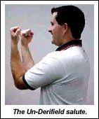
At the time, there were two Activator practitioners on the college faculty. One enjoyed discussing the matter with me, whereas the other (who in addition to his DC had a PhD in physics, or some such field) did not. Whenever I passed this latter in the hallways of the college, I would raise my arms in front of my chest, elbows bent at 900 (figure 1), and needle him: "If the lower legs are actually the same length, the feet must be held equally off the table. Pelvic subluxation can have nothing to do with it." One day, confronted one time too many by my un-Derifield salute (figure 1), he demanded that I desist, exclaiming he had seen with his very own eyes that the short leg often crossed over to become the long leg, and that was that. I asked him if that is what they had taught him of the scientific method in his PhD program: "I see it, therefore it happens!" Needless to say, we are still not friends.
Deconstructing a Moribund Derifield Model
When I moved over to teach at Palmer West in early 1989, I brought my obsession with the Derifield leg check with me. Working in the outpatient clinic, I developed a friendship with then-student and future Palmer West faculty member Dave Mullen (deceased). Out of our discussions emerged a model of the pelvic portion of the Derifield leg check, that led to a publication a little while later.3 Being very inexperienced at article writing at that time, I did not offer co-authorship to Dave (he most likely would have declined), or even acknowledge his contribution, but perhaps it is not too late to posthumously thank him for contesting so much of what I thought, since it is out of disagreement, not harmony, that the best elements of collaboration arise. Dave was a brilliant man, and left an indelible mark on Palmer West and also at the National Board of Chiropractic Examiners, where he worked on the Part IV test.
On the way toward developing a plausible Derifield pelvic model, it first became necessary to deconstruct the only prior model I had seen. Although I have never seen anything published on it, our college's main instructor in drop table methods used to show the students a device made of lengths of 1"x1" wood and hinges, that modeled how the knees-extended short leg could become the knees-flexed long leg, or stay short, as the case may be. The explanation depended on the fact that on the short leg side, the hip and knee would also be drawn cephalad. The only way to approximate the medial malleoli of the feet with the knees flexed required that the legs strike a different angle with the table top. The leg that is made more perpendicular to the table would then carry the foot on that side higher. If the short leg side is made more perpendicular to the table top, the finding would be D+, whereas if the long leg side is more perpendicular, the finding becomes D-.
Big problem. Depending on which leg the examiner decides to make more perpendicular to the table top, the D+/D- finding is random. To make matters worse, simple trigonometry can be used to calculate that a typical unleveling that might be seen in the knees-flexed position is unlikely to be clinically noticeable. Assuming a short leg of _" and a tibial of 20", and applying the Pythagorean theorem to triangle HTS, we find the difference in foot elevation, I, to be approximately a hundredth of an inch! So if Derifield lives, there will have to be some other way to make sense of the pelvic Derifield check.
What if the muscles in the anterior thigh compartment (quadriceps, sartorius) were stiffer, or less compliant, in one leg compared with the other? Then, in a prone patient lying with flexed knees, the weight of the lower leg will accomplish less compression of these stiffer anterior thigh muscles compared to the relatively softer muscles on the other side. Thus, the foot will be held relatively higher on the side of the stiffer anterior musculature.
This could occur on either the short or long leg side when the knees are extended. If it were to occur on the short leg side, we would get a D+ listing, a short leg crossing over to become long. If, on the other hand, this effect were to occur on the long leg side, then the long leg would remain long in the knees-flexed position (equivalent to the short leg remaining short), a D- listing.
It was not hard to put these hypotheses to an initial test, by creating a synthetic Derifield result, to see if firmer anterior thigh musculature would raise the foot.. The prone patient with knees flexed is asked to straighten the flexed knee against mild examiner resistance, increasing the tone of the quadriceps muscle, while the latter observes the relative positions of the feet. In performing this test, the foot on the contracted side rises, to an extent directly proportional to the magnitude of the patient's isometric effort. This constitutes a synthetic Derifield result, D+ if the knees-extended short leg is made to contract, or D- if the knees-extended long leg is made to contract.
Derifield-Positive, Derifield-Negative: A Biomechanical Hypothesis
Let us now bring this to bear on the Derifield leg check. Assume a state of pelvic torsion, with posterior innominate rotation on one side and relative anterior rotation on the other side. Although I don't have the space to properly develop the point, let us further assume a functional short leg on the posteriorly rotated innominate side. (That is a huge assumption, but I dare not go there in this article.)
In D+, weak thigh flexors (iliopsoas, rectus femoris and sartorius) would have allowed the short leg side innominate bone to subluxate posteriorly, inferiorly, and medially, resulting in what is commonly called a PI ilium. There is an unsuccessful effort on the part of the postural control mechanism to reduce pelvic torsion through increased thigh flexor tonus on the short leg side. Since this attempt to restore postural symmetry fails, the anterior thigh muscles are eccentrically contracted. Flexing the knees so as to elevate the legs further stretches the thigh flexors, which in turn further increases the stiffness of the sartorius and the rectus femoris component of the quadriceps group against the table. This may be due to stretch reflexes, a non-neurological firming due to tension, or both. The short leg crosses over and gets longer as the knees flex, because the anterior thigh compartment compresses less under the weight of the elevated leg. In short, we expect the rectus femoris and sartorius muscles to be relatively firmer on the side of the PI ilium, thus raising the foot relative to the contralateral foot, for the same reason that resisted knee extension, by tensing the quadriceps muscles, raises the foot.
In D-, thigh extensor contraction subluxates the innominate bone anteriorly and laterally, resulting in what is commonly called an AS ilium, and an assumed functional long leg on that side. There is a concentric contraction of the anterior thigh muscles. Flexing the knee in a prone patient, the AS side thigh flexors sustain increased stiffness, just as in the case of D+ (again: stretch reflexes, firming due to increased tension, or both), holding the ipsilateral foot relatively higher. Thus, the long leg gets longer as the knees flex, or, as it is usually stated, the short leg stays short.
In the larger scheme of things, a clear and dichotomous distinction may be drawn between the D+ and D- pelvic syndromes. Both clinical presentations involve pelvic torsion and chronic postural deviation, and yet arise in fundamentally opposite manners and manifest symmetric but opposite parameters. D+ signifies posterior innominate rotation allowed by eccentrically contracted, weak thigh flexors, and D- signifies anterior innominate rotation created by concentrically contracted, normal strength thigh flexors.
The Paper Sign
An orthopedic test emerges from the model, that either adds to or even replaces visualizing which foot is held higher off the table, either by looking at the shoes or the medial malleoli. The examiner places a piece of paper under both knees, about 3 inches in. The examiner assesses how much force is required to pull the paper out from under the knee, and then repeats the procedure on the other side. (It reminds us of Froment's sign, another paper test.)
On the side where the paper is easier to withdraw, the relatively increased quadriceps/sartorius tone has slightly raised the distal thigh, diminishing its contact against the paper on the table. It is reasonable to suppose that this paper sign might distinguish differences in foot elevation due to asymmetry in tibial lengths (negative paper sign) from those attributable to soft tissue asymmetry (positive paper sign). Of course, we can also do our best to rule out uneven tibial lengths by observing the knee elevation of the supine patient with knees flexed, and interpret the information conjointly.
A friend who practices the Activator technique once told me, that as an alternative to the paper sign, he had tried sliding an x-ray film under the knees of the prone patient with knees flexed, to see under which side the film would go more cephalad. I have not tried that out.
Derifield Epilegomenon
Yes, there are treatment implications, involving both adjustive procedures and soft tissue rehabilitation. Shortened soft tissues will need lengthening, and weak muscles will need strengthening (both directly, and by stretching tight antagonists). As one of the reviewers pointed out when I first submitted this model for publication,4 the Derifield pelvic leg check in essence identifies the side of the body wherein lies the primary pathology: the PI ilium on the short leg side of the D+ patient, and (usually) the AS side of the long leg side of the D- patient.
You, the reader, will have to take it from there. I have made up more than enough stuff in this article, a data-free, biomechanical model for the Derifield Pelvic leg check; and made a bloody mess of Occam's razor. Sorry.
References
Falltrick DR, Pierson DS. Precise measurement of functional leg length inequality and changes due to cervical spine rotation in pain-free subjects. J Manipulative Physiol Ther 1989;12(5):369-373.
Rhudy TR, Burk JM. Inter-examiner reliability of functional leg-length assessment. American Journal of Chiropractic Medicine 1990;3(2):63-66.
Cooperstein R, Bricker D, Jansen R. Detection of absolute and relative left and right leg displacements as a function of head rotation: the advantages of using a friction-reduced multi-segmented table. In: Cleveland III C, Haldeman S, editors. Conference Proceedings of the Chiropractic Centennial Foundation; 1995; Washington, DC: Chiropractic Centennial Foundation; 1995. p. 323-324.
Cooperstein R. The Derifield pelvic leg check: a kinesiological interpretation. Chir Tech 1991;3(2):60-65.
Dr. Robert Cooperstein, a professor at Palmer College of Chiropractic West, can be reached atwww.chiroaccess.com, or by e-mail atdrrcoop@aol.com.
Many relevant diagnostic signs are not performed deliberately by the examiner or by the patient at the examiner’s direction. They are observed as the patient reacts to their condition. Fortin’s finger sign, Minor’s sign, and Vanzetti’s sign are three examples of this principle.
On Feb. 1, 2024, two more legislators – John R. Carter (R-Texas) and Robert “Bob” Good (R-Va.) – co-sponsored the Chiropractic Medicare Coverage Modernization Act of 2023, bringing the co-sponsor total to 155, two more than the total achieved over the entire two-year congressional cycle in which identical legislation last appeared (2021-22).
While working at the Lovelace Medical Center in Albuquerque, N.M., one of the primary care physicians stated that imaging every patient prior to receiving chiropractic was not medically necessary. It was at that point I reconsidered the need for the imaging of every patient to avoid malpractice.