Cuboid subluxation is a poorly recognized condition, even though it is not uncommon. It has been described in the literature under various names: cuboid subluxation, cuboid syndrome, locked cuboid, dropped cuboid, cuboid fault syndrome or peroneal cuboid syndrome. It can be confused with a lateral ankle sprain, leading to mismanagement.
The American College of Sports Medicine defines a cuboid subluxation as a minor disruption or subluxation of the structural congruity of the cuboid articulations, particularly the calcaneocuboid joint, resulting in pain and impairment of joint function. Diagnostic imaging does not seems to help with establishing the diagnosis. The variability of the shape of the osseous structures and anomalies in the foot make it difficult to determine subtle joint space malalignments.
Ultrasound does offer some assistance in determining a true luxation, along with visualizing an intra-articular effusion, and allows for functional visualization. However, the primary diagnosis is made with clinical history and physical examination. Imaging is secondary and useful for ruling out other pathology such as fracture or fracture-dislocation.
Anatomy / Mechanics
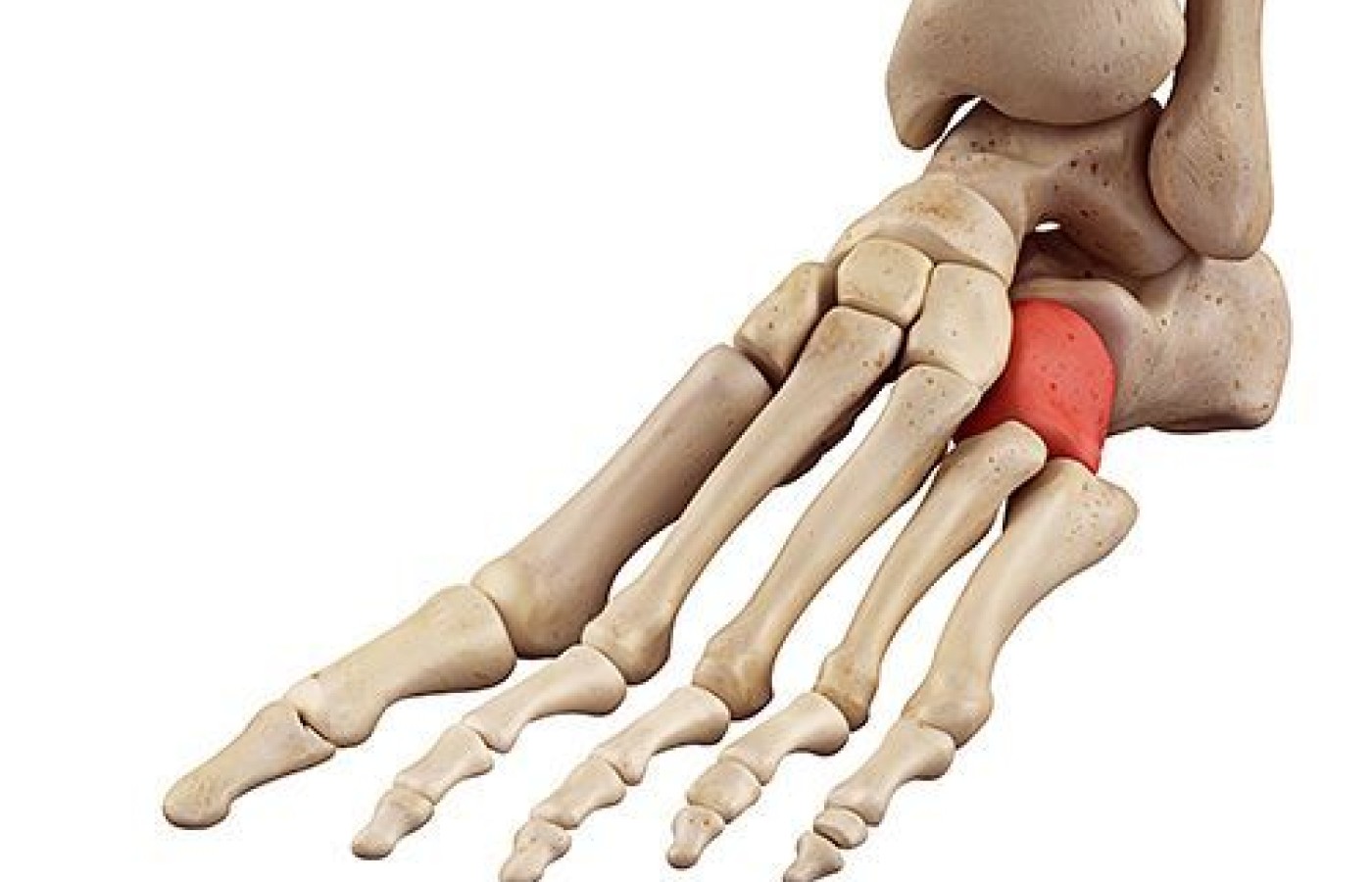
The cuboid is in the lateral midfoot, surrounded by the calcaneus posteriorly, the fourth and fifth metatarsals anteriorly, and the navicular and lateral cuneiform medially. Calcaneal-cuboid (CC) joint function is dependent on midtarsal joint mechanics and can be variable. The principal movement at the CC joint is medial-lateral rotation about an anterior-posterior axis, with the calcaneal process acting as a pivot.
The cuboid articulates with the lateral cuneiform and the navicular medially; and acts as the focal point of the lateral column for the transfer of force between the hindfoot and the forefoot. Gliding and rotation are the primary motions.
The peroneus longus tendon slings the cuboid as it runs on the undersurface of that bone into a groove, which is converted into the peroneal canal by the long plantar ligament. The tendon then crosses the sole of the foot obliquely and is inserted into the lateral side of the base of the first metatarsal bone and the lateral side of the medial cuneiform. Normal mechanics of the midtarsal joints are not fully understood, even to date.1
Symptom Portrait
The symptoms of cuboid subluxation resemble those of a ligament sprain. Pain is often diffuse along the lateral foot between the CC joint and the fourth and/or fifth cuboid-metatarsal joints, and may radiate throughout the foot. Depending on whether the injury is acute or chronic, a slight indentation over the dorsum of the cuboid may be present, or slight swelling on the plantar surface may be present in the region of the cuboid, along with erythema, edema or even ecchymosis.
Tenderness is often present along the peroneus longus tendon and at the cuboid groove. Range of motion of the ankle and/or foot, both active and passive, may be decreased due to pain. Resisted ankle / foot eversion or inversion will elicit pain. Walking may be affected; an antalgic gait is common. Hopping will increase symptoms.
Possible Causes and Mechanisms of Injury
Several causes have been proposed for cuboid subluxation, including excessive pronation, overuse and inversion ankle sprains. The precise pathomechanic mechanism is unclear; it is thought to be caused from forceful eversion of the cuboid while the calcaneus is inverted, with resultant disruption of CC joint congruity. The peroneus longus may play a role in the development of cuboid subluxation, since this muscle imparts an eversion moment on the cuboid.
Mechanisms of injury include acute, traumatic and chronic, as in overuse. Incidence is quite common in ballet dancers – as high as 17 percent.2
Plantar flexion and inversion ankle sprains account for most traumatic cuboid subluxations. This form of injury is more common in male dancers and involves landing from a big jump or a series of big jumps with a forceful foot pronation.
The overuse form of injury is more common in female dancers, in particular pointe work. Entry into the en pointe position initially causes a dorsiflexion of the tarsometatarsal articulations; upon reaching full ankle plantar flexion, if the dancer's balance carries her farther, an opposite plantar flexion moment is experienced at the tarsometatarsal joints.
Forceful contraction of the peroneus longus or tightening of the tendon passively during forceful foot plantar flexion, inversion and adduction forces the cuboid into a closely packed position; one could describe it as jamming it into a plantar medial displacement. It is these repetitive forces during dance that destabilize the midfoot and predispose a dancer to cuboid subluxation.3
All these mechanical forces can injure the CC joint, possibly resulting in ligament laxity. Of course, one does not have to be ballet dancer to have this injury occur; gymnasts and even runners have a higher incidence of this type of injury than the normal population.
Differential Diagnosis
Because of the difficulty in diagnosing cuboid subluxation, differential diagnosis of lateral foot pain should include some of the more common injuries of the lateral foot, including fracture or dislocation of the cuboid, calcaneus, or fourth or fifth metatarsals. Other possible causes of lateral foot pain include lateral midfoot sprain, peroneus longus pathology, arthritis, tarsal coalition, sinus tarsi syndrome and symptomatic os peroneum syndrome.
Radiographic evaluation can be performed primarily to rule out these other entities. Cuboid subluxation also can be misdiagnosed as a lateral ankle sprain or can be present in conjunction with a lateral ankle injury. If CC joint pain persists after lateral ankle symptoms have subsided, a cuboid subluxation should be highly suspected.
As previously noted, most imaging modalities including CT and MRI are not helpful in establishing the diagnosis of cuboid subluxation. Ultrasound evaluation may, however, be helpful in diagnosing traumatic cuboid or midfoot injuries.
Musculoskeletal ultrasound has not been widely available until recently; this modality allows for confirmation of the diagnosis and ability to assess the reduction – and also the possibility to assess other soft-tissue injuries, such as peroneal tendon pathology.
Functional studies can be performed to evaluate stability of the articulations. Ultimately, however, the history and physical exam are the most important tools for determining the diagnosis.
There are no definitive, validated diagnostic tests for cuboid syndrome, but two clinical maneuvers have been described: 1) the midtarsal adduction test; and 2) the midtarsal supination test.4
Manipulation Options
The most appropriate treatment for cuboid subluxations, both acute and chronic injuries, is manual manipulation. The more acute, traumatic injuries, especially those associated with swelling and ecchymosis, should be treated with careful manipulation to avoid further injury to damaged ligaments and the peroneus longus tendon.
Less significant acute and subacute injuries involving subluxation without extensive soft-tissue damage respond well and may need to be adjusted a few times over a few days or weeks. Chronic injuries may require repeat manipulations and possible external fixation in a cast or boot. Taping is also recommended initially in either case; along with icing after adjustments to reduce any swelling.
The goal when manipulating is to reduce a plantar and medially subluxated cuboid. The most common, safest and easiest reduction maneuver is the cuboid squeeze. The patient is prone and the foot and ankle are in maximum plantar flexion. The clinician applies mild axial distraction with both hands while using both thumbs overlapping on the cuboid, pressing the cuboid in the dorsal and lateral direction.
An alternative method is the cuboid whip, which is performed with the affected foot hanging freely in the air with the knee flexed to 90 degrees. Holding the foot in the similar position as squeeze technique, the clinician gently swings the knee, ankle and foot in an arc in the sagittal plane; once guarding is minimal and muscles / tendons relax, a dorsal-lateral high-velocity, low amplitude adjustment can be made.
Manipulation is usually effective for acute and subacute plantar subluxations. Newell and Woodle report that a cuboid subluxation present for less than one week will respond with 1-2 adjustments. If symptoms are present for only a month, most will respond with 2-4 adjustments. Symptoms present for six months require repeated treatment and evaluation for six months or more.5
Dorsal subluxations of the cuboid are rare, but will respond to manipulation as well. The technique for adjusting the dorsal subluxation can be performed with the patient seated and foot flat on the table or floor, stabilizing their ankle (primarily the distal fibula) as you apply traction to the fourth and fifth metatarsals with one hand, and a downward thrust to the dorsal surface of the cuboid with the other hand. An audible often will be heard with a relief in symptoms.
Cuboid adjustments may hold without external support, but if resubluxation occurs, support should be considered. Icing after adjustment is highly recommended.
The biggest challenge with cuboid subluxations is for the patient to get proper treatment in a timely manner. Many patients initially are seen by clinicians who are not aware of this condition, leading to imaging studies and useless treatments – some that even aggravate the condition. Hopefully as this condition becomes more recognized as a subluxation, more patients will receive appropriate chiropractic care.
References
Durall CJ. Examination and treatment of cuboid syndrome: a literature review. Sports Health, 2011;3(6):514-519.
Marshall P, Hamilton, WG. Cuboid subluxation in ballet dancers. Am J Sports Med, 1992;20:169-75.
Adams E, Madden C. Cuboid subluxation: a case study and review of the literature. Current Sport Med Rep, 2009;8(6):300-307.
Jennings J, Davies GJ. Treatment of cuboid syndrome secondary to lateral ankle sprains a case series. J Orthopedic Sports Phys Ther, 2005;35(7):409-415.
Newell SG, Woodle A. Cuboid syndrome. Physical Sports Med, 1981;9:71-6.
Many relevant diagnostic signs are not performed deliberately by the examiner or by the patient at the examiner’s direction. They are observed as the patient reacts to their condition. Fortin’s finger sign, Minor’s sign, and Vanzetti’s sign are three examples of this principle.
Spearheaded by burgeoning scientific and clinical research literature, psychedelics have reached a level of media coverage and popular interest that has not been seen for over half a century. By “psychedelics,” we are referring to the unique class of substances that includes psilocybin (the active compound found in so-called “magic mushrooms”), LSD, dimethyltryptamine (DMT), ayahuasca, 5-MeO-DMT, and mescaline – each of which occurs in the natural world (except for LSD, which is a semi-synthetic compound).
While working at the Lovelace Medical Center in Albuquerque, N.M., one of the primary care physicians stated that imaging every patient prior to receiving chiropractic was not medically necessary. It was at that point I reconsidered the need for the imaging of every patient to avoid malpractice.