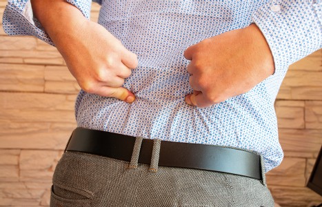
Many relevant diagnostic signs are not performed deliberately by the examiner or by the patient at the examiner’s direction. They are observed as the patient reacts to their condition. Fortin’s finger sign, Minor’s sign, and Vanzetti’s sign are three examples of this principle.
Treat Every Patient as an Athlete
Frontal-plane movement pattern dysfunction can set the stage for musculoskeletal injury. Frontal-plane stabilization is essential during the normal activities of daily living: think single-leg stance and gait cycle. Add asymmetrical loads such as carrying a suitcase, groceries, kids, a purse, etc., and the system gets challenged even more.
Athletes are extremely vulnerable to frontal-plane injury because of dynamic load variables during the chaos of competition. But in reality, everyone is an athlete. Why? Because life is chaos. You never know when, where or how something is going to come at you. Nike co-founder Bill Bowerman said, "If you have a body, you are an athlete." What a powerful statement. It's our obligation to treat every patient as an athlete and assess their ability to stabilize in every plane of motion: sagittal, frontal and transverse. Optimal mobility can only be achieved when the body has a stable foundation.
Lateral Flexion and the QL

Lateral flexion is frontal-plane movement. The quadratus lumborum (QL) muscle is a primary driver of lateral flexion and culprit in most lower back pain syndromes. How often do you find one or both of them as a problem? Treatments such as soft-tissue manipulation, stretching, ultrasound, muscle stimulation, laser therapy, strengthening, etc., are relentlessly thrown at the QL. All are great options for treating the pain; however, we need to ask ourselves, Why and how is the quadratus lumborum dysfunctional? Is it painful and weak (inhibited) or painful and strong (facilitated)? Is it compensating for something else and working too much, or not doing enough during movement demands? The answer to those questions will dictate the course of action taken in restoring its proper function in lateral flexion movement patterning.
What Is the Action of the Quadratus Lumborum?
- Unilaterally elevates the pelvis
- Laterally flexes the vertebral column to the same side
- Assists to extend the spine
- Bilaterally works to fix the last rib during forced inhalation / exhalation
The opposite-side quadratus lumborum does the antagonistic muscle action. The QLs must have a functional relationship with each other. When one is in a concentric action, the other must eccentrically control the motion.
What Other Muscles Work With the QL to Laterally Flex the Spine?
- Lumbar paraspinals
- External oblique
- Internal oblique
- Psoas major (assists)
- Latissimus dorsi (assists)
What happens if a QL is inhibited (neurologically down-regulated)? The nervous system must use more of another muscle in a pattern to assist movement (commonly synergistic) such as the lumbar paraspinals; or it may overuse an antagonistic muscle, otherwise known as a functional opposite. A common culprit in this compensation pattern is the opposing quadratus lumborum.
For example, an inhibited right quadratus lumborum may cause facilitation (neural up-regulation) to the right lumbar paraspinal (synergistic pattern) and/or the left quadratus lumborum (antagonistic pattern). The patient might experience right-paraspinal or left-sided QL pain resulting from a dysfunctional right QL.
What happens if the internal and external oblique muscles are inhibited? (This is a very common pattern.) Again, the nervous system will then rely on another muscle in the pattern to take over the workload. For example, if the right internal and external obliques are inhibited, the right quadratus lumborum or lumbar paraspinals may become facilitated, causing pain. In order to alleviate the QL facilitation and pain, you must activate the obliques.
How Do You Determine Which Pattern to Correct? Assess
Observe static posture: Is there a lateral bend to one side? Is there a closing down of the space between the rib cage and pelvis on one side? Is one shoulder higher than the other? These may indicate a shortening in one QL / oblique compared to the other.
Palpation: What is the muscle tone of the lateral flexors (QL / obliques / paraspinals / latissimus)? How do they compare to each other and the functional opposite side? Are they hypertonic or hypotonic? Are there trigger points in the muscles?
Movement: Observe the patient in a single-leg stance. Do they present with Trendelenburg's sign? Does the pelvis drop on the side of the leg in the air, or does the patient over-hike the pelvis to compensate?
Next, instruct the patient to perform the standing side-bend test: feet shoulder-width apart and slight bend in the knees, hands over head. Have them bend to the right and then to the left. What is the quality of their movement: slow and controlled or fast and loose? Is there trepidation in the movement? Do they extend or rotate the spine more than side bend? Do they shift the pelvis in a frontal-plane motion?
Watch the neck during this maneuver as well. A common compensation in side-bending is to laterally flex the neck instead of the lumbar spine. That tells you they are using the neck to drive the pattern, which may result in neck pain.
Muscle Testing the QL
Muscle testing is a great way to compare one QL to the other. You can do this with the patient in a supine or standing position. Supine position: Stand on one side of the patient (for this explanation, let's choose the right side). Have patient move both legs to the left at least 6 inches. Stabilize the outside of the right hip with your left hand. Take your right hand / forearm under the ankles of both legs and grab outside of the left ankle. Try pulling the legs back to center as the patient resists. Cue the patient to resist your motion.
Pay careful attention to whether the patient holds their breath or grabs the table for stability. Do their legs move easily back to center or do they lock in and hold? If they cannot maintain the position, the left QL is inhibited.
Now walk to the left side of the body and have the patient bring their legs to the right 6 inches. Stabilize the outside of the left hip with your right hand and place the left hand under the ankles, holding onto the right ankle. Pull legs back toward center. Can they resist the motion? This is assessing the right QL; the side that tests weak is the inhibited side.
A progression is to have patient hold the table in a supine position. Move their legs 6 inches to one side. Lift legs off the table near the ankle. Repeat the test, but add more pressure. Do they lock and hold, or do the legs weaken?
Muscle test in this sequence: Side 1. Side 2. Side 1. For example: Test right QL (1); then test left (2); then test right again (1). You are comparing relationships. They should remain strong and locked during all parts of this sequence.
With the patient in a standing position, feet shoulder-width apart, slight bend in the knees and hands over head, stand on their right side. Place right hand on the latissimus muscle just below the armpit. The left hand goes on the outside of the left hip. Try pressing the patient back to center with your right hand. They should be able to maintain the position. This is testing general right lateral flexion patterning.
Switch sides and hand positions. Compare sides. Now repeat the test, but do not hold the opposite hip. Do they lose balance? Is it less stable? If so, the side they are flying to is weaker / inhibited.
What happens if both sides test weak? Think logically. This indicates massive instability in the system and is often associated with a subluxated lumbopelvic hip complex (LPHC). You must adjust the LPHC and then add stability correctives.
What If You Find an Asymmetry?
You must activate and strengthen the weaker / inhibited muscles in conjunction with releasing the ones that are overcompensating. For example: If the right QL is weak and the left is strong, do modality work to both sides and then strengthen the right. What's the best way? Simply use the muscle in its intended pattern. Side bend and extend or do a low-level stabilization side-plank to tolerance. What if the right QL is inhibited and the right external oblique facilitated? Release both and then activate the right QL with a knees-bent side plank.
Muscles that function together and share the load make for efficient movement patterning and less energy expenditure. The ability to absorb, direct / disperse, generate and release force is fundamental to stability and injury prevention. Muscular inhibition will cause kinetic-chain dysfunction and energy leaks, resulting in compensation. In your next assessment, focus on frontal-plane stabilization assessment and correction to reduce the chances of musculoskeletal pain arising from these compensations.
Editor's Note: Check out this video demonstration by Dr. Nickelston showing how to muscle test for QL dysfunction.
Resources
- Chaitow L, Walker-DeLany J. Modern Neuromusculoskeletal Techniques. New York: Churchill Livingstone, 1996.
- Cook G. Movement: Functional Movement Systems: Screening, Assessment, & Corrective Strategies. Aptos, CA: On Target Publications, 2010.
- Page P, Frank C, Lardner R. Assessment and Treatment of Muscle Imbalance: The Janda Approach. Champaign, IL: Human Kinetics, 2010.
- Vleeming A, Mooney V, Stoeckart R. Movement, Stability & Lumbopelvic Pain: Integration of Research and Therapy. Edinburgh: Churchill Livingstone Elsevier, 2007.
- Weinstock D. NeuroKinetic Therapy: An Innovative Approach to Manual Muscle Testing. Berkeley, CA: North Atlantic, 2010.


