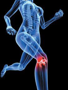
A Nonsurgical Approach for Treating Meniscus Injury
Warren Hammer, MS, DC, DABCO
Patients occasionally enter the office with a torn meniscus of the knee. In these cases, it is important to determine if they could respond to conservative care. Meniscal tears may be either traumatic or degenerative, and degenerative tears are closely associated with osteoarthritis. Based on symptomatology, examination and age, one might consider a degenerative meniscal tear from a plain X-ray, but acute tears do not have any specific radiographic findings.1
While magnetic resonance imaging (MRI) is considered the best method for visualizing the knee meniscus, the tear appearing on MRI has no significant basis unless it is based first on the history and physical examination. Between 36-76 percent of asymptomatic patients show tears on MRI and the percentage greatly increases with age. Asymptomatic patients older than age 65 show meniscal tears at a 67 percent rate, and tears are prevalent in 86 percent of patients with symptomatic osteoarthritis.2 Since asymptomatic meniscal tears are common, it is essential that a practitioner be certain that the meniscal tear is the source of the patient's pain.
One of the most important factors with conservative vs. surgical care is the location of the tear with regard to meniscal vascularity, since the areas of the meniscus with the most vascularity have the greatest ability to heal. The periphery of the menisci is where the blood supply originates (perimeniscal capillary plexus), which derives its supply from the outer medial and lateral geniculate arteries.
Only the peripheral 25-30 percent of the meniscus is vascularized,1 leading to a division of the meniscus with regard to its circulation. The outer third is called the red-red zone, the middle third is called the red-white zone and the remainder of the internal zone (adults) is called the white-white zone. In the red-red zone, bleeding can result in the formation of fibrovascular scar tissue and attract anabolic cells due to cytokines released during the inflammatory phase. The red-white zone has less vascularity and healing potential is therefore less. The white-white zone receives nutrition from synovial fluid by passive diffusion, which can be stimulated by knee joint motion, but since a healing response is not created, the prognosis is poor regarding surgical repair in this zone.
Healing is influenced by the pattern of the tear and the type of vascularity. Longitudinal tears heal better than radial tears. Simple tears heal better than complex tears. Traumatic tears have higher healing rates than degenerative tears, and acute tears heal better than chronic tears.3
Of the many tests used to diagnose a meniscal tear, tenderness at the medial joint line scores highest in terms of accuracy (76-86 percent), the Apley grinding test 46 percent, painful hyperextension 43 percent, Steinmann I sign 42 percent, and McMurray test 35 percent. Most meniscus tests attempt to trap abnormally mobile fragments of the menisci between the femur and the tibia, causing either pain or clicking; but the use of joint-line tenderness in patients with both a meniscus tear and an acute anterior cruciate ligament tear has been shown to be completely unreliable. Joint-line tenderness is most reliable when the tenderness is localized to the posteromedial or posterolateral corner of the knee, since anterior joint-line tenderness is usually present with patellofemoral disease.4
A simple "squat test," whereby the patient raises and lowers themselves from a crouched position, is more indicative of a patellofemoral problem as long as the squat is less than 90 degrees flexion. In 90 degrees knee flexion, 85 percent of the joint load is transmitted through the menisci.4
A particular soft-tissue method I have found beneficial over the years for meniscal tears relates to the treatment of peripheral tears. Free nerve endings of the meniscus are only found on the peripheral one-third, and since palpation of the meniscus at the joint line seems to be the most acceptable test for determining a meniscal lesion, you might assume that the lesion may be more peripheral where the circulation is most prominent.
For the medial joint-line tender area, if it is the right knee, flex the patient's knee 90 degrees and externally rotate the foot and tibia to open up the medial space. Externally rotating the foot also puts pressure on the medial meniscus tear from the medial femoral condyle. Internally rotate the foot and tibia for a lateral tear. Use friction massage with your finger or Graston Technique over the area until you feel a marked decrease in tissue density. (This might take up to 5 minutes.) Do not repeat this treatment for at least five days.
Pain during knee flexion implicates the posterior horns. Pain with extension implicates the anterior horns. Internal rotation tests the lateral meniscus while external rotation tests the medial meniscus.
Manipulation may be performed within the first 24 hours of acute locking (swelling may prevent manipulation) or in chronic locking. Mennell describes a manipulation for a medial-locked torn meniscus as follows.4 He states that in all his years of treating meniscal locking by manipulation, he has never had to treat a lateral lock.
Standing on the supine patient's left side, flex the patient's left knee approximately 110 degrees.
Put your left forearm over the patient's lower tibia and medial malleolus, grasp the calcaneus with the hand and then externally rotate the foot (the entire procedure may also be attempted with the foot internally rotated).
With the right hand, steady the knee with a minor valgus stress. It is important that the tibia remain in a sagittal neutral position (no varus or valgus) during the entire manipulation.
Fully flex the knee, literally kicking the knee into the buttock.
If locking is not immediately reduced, the patient should be referred to an orthopedist.
Estimation of the severity of the lesion, the age of the patient, the degree of knee instability, and the patient's occupation all have significance in determining whether conservative treatment may be attempted. Prolonged loss of knee extension, chronic severe pain, locking, and swelling are definite indicators for possible surgery. An acute injury in young patients (usually in their 20s) should always make one suspect more than just the isolated meniscus lesion. Usually there is pain, swelling, giving way and locking. Often the meniscus tear is associated with an MCL or ACL lesion.
In an athlete, an acute meniscus lesion usually prevents the individual from walking off the field unaided. (This is not the case with an isolated ligamentous injury.) The patient usually complains of a giving way at the time of injury. The meniscus may displace, causing an immediate loss of extension. Patients with medial meniscal tears create a knee locking at 10-30 degrees of flexion, while laterally displaced meniscal tears lock in greater degrees of flexion, especially posterior tears at more than 70 degrees.
Pain at extreme knee extension is affecting the anterior horn, while pain at extreme knee flexion is affecting the posterior horn. The pain in a medial meniscus injury is more often in the posterior medial or medial joint line and is rarely localized anteromedially. Lateral meniscus joint line pain is more often midlateral than posterolateral.4
A degenerative tear of the meniscus in an older patient (40 years or older) is more likely to be an isolated lesion. Older individuals may develop a tear for no apparent reason or feel it as they arise from a chair. If they develop chronic symptoms, they may present with thigh atrophy and weakness associated with pain, effusion, and giving way.
An isolated meniscus lesion will develop mild effusion gradually over a few days, compared with the almost immediate swelling of an anterior cruciate lesion, although swelling can be rapid in a meniscus tear if it occurs in the peripheral vascular zone. If the swelling occurs a day or so later, the tear is probably in the nonvascular central meniscal area. Immediate locking (loss of knee extension) may occur, especially if a bucket-handle tear (longitudinal type) occurs.
Failure to extend the knee may be due to eventual effusion (hamstring spasm and pseudo-locking), so it is important to question the patient to find out whether it was possible to extend the knee fully immediately after the injury. Rarely, a posterior vertical tear of the lateral meniscus will cause a locking in full flexion.4 The majority of the time, knee locking occurs in extension. Of course, rehabilitation, stretching and strengthening should be included with a conservative approach.
References
Mazak TG, Fabricant PD, Wickiewicz TL. Indications for meniscus repair. Clinics in Sports Med, 2012;31(1):1-14.
Greis PE, Bardana DD, Holmstrom MC, et al. Meniscal injury: basic science and evaluation. J Am Acad Orthop Surg 2002;10(3):168-76.
McCarty E, Marx RG. Meniscal tears in the athlete. Operative and nonoperative management. Phys Med and Rehab Clin North Amer, 2000;11(4):867-878.
Hammer W. Functional Soft Tissue Examination and Treatment by Manual Methods, 3rd Edition. Jones & Bartlett: Sudbury, MA, 2007.
Many relevant diagnostic signs are not performed deliberately by the examiner or by the patient at the examiner’s direction. They are observed as the patient reacts to their condition. Fortin’s finger sign, Minor’s sign, and Vanzetti’s sign are three examples of this principle.
On Feb. 1, 2024, two more legislators – John R. Carter (R-Texas) and Robert “Bob” Good (R-Va.) – co-sponsored the Chiropractic Medicare Coverage Modernization Act of 2023, bringing the co-sponsor total to 155, two more than the total achieved over the entire two-year congressional cycle in which identical legislation last appeared (2021-22).
While working at the Lovelace Medical Center in Albuquerque, N.M., one of the primary care physicians stated that imaging every patient prior to receiving chiropractic was not medically necessary. It was at that point I reconsidered the need for the imaging of every patient to avoid malpractice.