The Ischial Spine: A Radiographic Sign for Acetabular Retroversion
Deborah Pate, DC, DACBR
Primary osteoarthritis of the hip appears to have a structural basis. It was hypothesized that acetabular retroversion could be associated with osteoarthritis of the hip by Murray1 and Stulberg2 in the mid '60s and early '70s. They were the first to describe this association between abnormal hip anatomy and its possible role in the development of osteoarthritis (OA).
In the past few years, femoroacetabular impingement (FAI) has become recognized as a cause of OA in adults. Much has been written about this entity and most clinicians are now aware of FAI. Because of its association with FAI, it's important to recognize acetabular retroversion when it presents clinically.
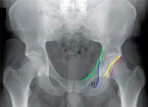
In the normal hip, the acetabular opening is anterior. In the retroverted condition, the superior aspect of the acetabulum is tilted posteriorly. On the AP X-ray in a normal acetabulum, the anterior rim of acetabulum should always project medial to the posterior wall. In cranial acetabular retroversion, the anterior rim will project lateral to the posterior wall in the superior aspect of the acetabulum.
Figure 1: Edge of anterior acetabular wall (yellow line) is medial to the edge of posterior wall (red line), even in superior aspect of acetabulum. Note that these lines never cross. Green line is rim of the pelvis, iliopectineal line. Blue line is acetabular teardrop.
One of the subtle anatomic abnormalities linked with femoroacetabular impingement is acetabular retroversion. It is now recognized that this anatomic abnormality results in cartilage degeneration secondary to femoroacetabular impingement, resulting in OA of the hip.3-4 Retroversion of the acetabulum is described as an abnormal morphologic feature involving a posteriorly oriented cranial acetabular opening in the sagittal plane, which is seen as the crossover sign (COS) on standard radiographs. The COS as seen on standard AP radiographs is indicates the presence of acetabular retroversion. Jamali5 confirmed that COS indicates retroversion of the acetabulum, which primarily affects the cranial portion of the acetabulum.
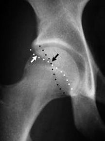
Figure 2: Edge of anterior wall (white arrow) is medial to the edge of posterior wall (black arrow) in inferior segment of the hip, but in the superior segment, this relationship reverses. This leads to the "crossover sign."
Interpreting the AP pelvic radiographs, however, can be difficult, and observing the anterior and posterior margins of the acetabulum is not always possible due to technique and quality of the radiograph. It has been observed that along with the crossover sign, the ischial spine is commonly projected into the pelvic cavity. This finding is easier to observe even when the crossover sign is not visible. Kalverer6 confirms that the crossover sign and the prominence of the ischial spine into the pelvis are reliable findings confirming the presence of acetabular retroversion.
Retroversion of the acetabulum is also associated with an externally rotated hemipelvis, which leads to protrusion of the ischial spine into the pelvis on the AP projection. COS and PRIS signs can be used to evaluate patients with early symptomatic femoroacetabular impingement and can also be used to detect hips at risk when the condition is bilateral, but only one side is symptomatic. The PRIS can be used even in pediatric cases because the acetabular walls are not ossified, so the contours are not visible on an AP radiograph.
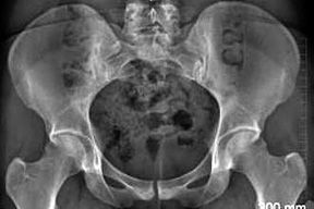
Figure 3: Ischial spine is projecting (PRIS) into the pelvis on both sides.
A recent study by Siebenrock, et al.,7 nicely demonstrates that hip impingement and decreased hip internal rotation in adolescent athletes exist more frequently compared to non-athletes. This suggests an important developmental etiology of hip impingement based on exposure to stresses during skeletal growth. Being able to recognize radiographic signs and clinical symptoms of FAI can help prevent the secondary changes associated with OA, especially in the younger patient. Further research is needed to determine the exact timing in skeletal age and features of repetitive stress that lead to pathologic changes.
References
Murray RO. The aetiology of primary osteoarthritis of the hip. Br J Radiol, 1965;38:810-824.
Stulberg SD, Cordell LD, Harris WH, Ramsey PL, MacEwen GD. Unrecognized childhood hip disease: a major cause of idiopathic osteoarthritis of the hip. In: Amstutz HC, ed. The Hip: Proceedings of the Third Open Scientific Meeting of the Hip Society. St. Louis, MO: CV Mosby; 1975:212-228.
Ganz R, Parvizi J, Beck M, Leunig M, Notzli H, Siebenrock KA. Femoroacetabular impingement: a cause for osteoarthritis of the hip. Clin Orthop Relat Res, 2003;417:112-120.
Giori NJ, Trousdale RT. Acetabular retroversion is associated with osteoarthritis of the hip. Clin Orthop Relat Res, 2003;417:263-269.
Jamali AA, Mladenov K, Meyer DC, Martinez A, Beck M, Ganz R, Leunig M. Anteroposterior pelvic radiographs to assess acetabular retroversion: high validity of the 'cross-over' sign. J Orthop Res, 2007;25:758-765.
Kalberer F, Sierra RJ, Madan SS, Ganz R, Leunig M. Ischial spine projection into the pelvis: a new sign for acetabular retroversion. Clin Ortho & Rel Res, 2008;466(3):677-683.
Siebenrock KA, et al. The cam-type deformity of the proximal femur arises in childhood in response to vigorous sporting activity. Clin Ortho & Rel Res, July 15, 2011.
For more information on femoroacetabular impingement (FAI), review: Tannast M, et al. Femoroacetabular impingement: radiographic diagnosis - what the radiologist should know. AJR, June 2007;188(6):1540-1552.
Many relevant diagnostic signs are not performed deliberately by the examiner or by the patient at the examiner’s direction. They are observed as the patient reacts to their condition. Fortin’s finger sign, Minor’s sign, and Vanzetti’s sign are three examples of this principle.
Spearheaded by burgeoning scientific and clinical research literature, psychedelics have reached a level of media coverage and popular interest that has not been seen for over half a century. By “psychedelics,” we are referring to the unique class of substances that includes psilocybin (the active compound found in so-called “magic mushrooms”), LSD, dimethyltryptamine (DMT), ayahuasca, 5-MeO-DMT, and mescaline – each of which occurs in the natural world (except for LSD, which is a semi-synthetic compound).
While working at the Lovelace Medical Center in Albuquerque, N.M., one of the primary care physicians stated that imaging every patient prior to receiving chiropractic was not medically necessary. It was at that point I reconsidered the need for the imaging of every patient to avoid malpractice.