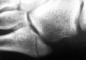
Fractures of the proximal fifth metatarsal are often separated into two types: those involving the tuberosity and those involving the diaphysis just distal to the tuberosity. It is important to appreciate this distinction because these two fractures respond differently to treatment. Fractures involving the tuberosity treated conservatively usually heal clinically within three weeks and radiographically within eight weeks. Fractures through the proximal diaphysis just distal to the tuberosity - the classic Jones fracture (Figure 1) - do not heal as quickly, often taking more than 11 weeks. The Jones-type fracture may not respond to conservative treatment, with delayed union or non-union occurring 40 percent of the time and requiring surgical intervention.1
Fig. 1: Jones fracture. Note the perpendicular fracture line.
The fifth metatarsal is divided into the base, the tuberosity, the shaft (diaphysis), the neck and the head. The tuberosity, also termed the styloid process, is a bony prominence just distal to the base. Both the peroneus brevis and peroneus tertius have insertions adjacent to this prominence. The plantar fascia also has a focused insertion on the proximal tip of the fifth metatarsal. The distal metaphysis tapers to the tubular diaphysis of the fifth metatarsal. Fractures commonly occur at this metatarsal shaft, within 1.5 cm of the tuberosity.
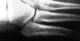
Avulsion fractures of the tuberosity are the most common fractures involving the proximal fifth metatarsal. Avulsion fractures occur proximal to the metaphyseal-diaphyseal junction. (Figure 2) If not displaced or comminuted, these fractures uniformly heal well with conservative treatment within three weeks.
It is important to differentiate avulsion fractures from an apophysis and from a fracture at the metaphyseal-diaphyseal region, as the treatment and healing time can be vastly different. The avulsion fracture is usually perpendicular to the long axis of the fifth metatarsal and the fracture is through the tuberosity and does not involve the metaphyseal-diaphyseal junction. In contrast to fractures, an apophysis has an oblique orientation and the lucent line between the metatarsal and apophysis should align parallel to the diaphysis. (Figure 3)
Fig. 2: Avulsion fracture. Note the oblique fracture line through the tuberosity.
The development of a secondary center of ossification at the proximal end of the fifth metatarsal appears in girls ages 9 to 11 and boys ages 11 to 14, appearing as a fleck of calcification adjacent to the fifth metatarsal shaft on plain films. The apophysis has an oblique orientation, with the radiolucency aligned parallel to the fifth metatarsal diaphysis. (Figure 4) The orientation of the radiolucency is crucial to differentiating this normal finding from an acute fracture.
There is, however, a self-limiting disorder: apophysitis (Iselin's disease), which is seen in children and spontaneously resolves with completion of growth. Symptoms include complaints of localized pain on activity that resolves with rest. Rarely, associated ecchymosis and edema can be present. Physical examination reveals tenderness at the fifth metatarsal base. Radiographic findings include irregular apophyseal density and shape. Treatment is primarily limitation of activity.
Fig. 3: Location and orientation of fracture determines type of fracture.
Differentiating from an avulsion fracture can be very difficult or impossible if the apophysis has not fused and is comminuted. In these cases, treatment should be the same as if a fracture were present. Again, generally these problems respond to conservative treatment including rest, an elastic bandage, or partial weight-bearing with crutches if the symptoms are severe.
Fig. 4: Apophysis of the fifth metatarsal.
Of course, we can't forget the accessory ossicles that are so common in the foot, which may also be confused with avulsion fractures. The os vesalianum is quite rare and may be found next to the peroneus brevis insertion. The more common os peroneum can be located at the lateral border of the cuboid within the substance of the peroneus brevis tendon. Each ossicle can be radiographically differentiated by its smooth appearance and generally is not symptomatic; in contrast, avulsion fractures appear to have "scalloped" edges. However, there are accessory ossicles that are symptomatic, which can confuse the diagnosis.
While differentiating an avulsion fracture from an apophysis and from accessory ossicles may be difficult, the Jones fracture has very specific criteria, making it easier to identify. A fracture involving the metaphyseal-diaphyseal region within 1.5 cm distal to the tuberosity of the fifth metatarsal is considered a Jones fracture. Torg and colleagues developed the classification system based on historical and radiographic criteria.2,3(See table below)
Torg's Classification System for Proximal Fifth Metatarsal Fractures (Within 1.5 cm of the Tuberosity)
Classification (Type)
Radiographic Appearance
Type 1
No intramedullary sclerosis; fracture line with sharp margins and no widening; minimal cortical hypertrophy; and minimal evidence of periosteal reaction to chronic stress.
Type II, delayed
Fracture line that involves both cortices with associated periosteal bone union; widened fracture line with adjacent radiolucency related to bone resorption; and evidence of intramedullary sclerosis.
Type III, nonunion
Wide fracture line; periosteal new bone and radiolucency; and complete obliteration of the medullary canal at the fracture site by sclerotic bone.
It is important to determine the type of fracture because before the development of Torg's classification system, high rates of nonunion were reported in patients with Jones fracture. It has been postulated that these nonunions were due to disruption of the vascular supply, which enters the bone in the metaphyseal-diaphyseal region. If, however, the fracture is properly classified as type I or type II, most patients will respond to conservative treatment; the possible exception is elite athletes, whose healing time is crucial to their livelihood.
Fig. 5: Jones type 1 fracture.
Type I fractures are treated with a non-weight-bearing, short-leg cast for six to eight weeks, with progressive ambulation after cast removal. Type II fractures may also be treated with a non-weight-bearing cast, but a prolonged period may be required until union is achieved. In competitive athletes, these fractures are usually treated operatively, either with medullary curettage and inlay bone grafting or intramedullary screw fixation. Type III fractures should be treated operatively.
Complications associated with surgical fixation (intramedullary screw) include persistent pain at the base of the fifth metatarsal requiring screw removal and shoe modifications. Delayed union, nonunion or refracture are also complications associated with surgical fixation.
Fig. 6: Jones type II fracture. Note the medullary sclerosis.
Although fractures of the proximal fifth metatarsal can be confusing as far as categorization, it's always best to treat as if there is a fracture. Special imaging such as MRI and bone scans will confirm the presence of a fracture and may be necessary if the patient doesn't respond to treatment. Remembering that the location of the fracture site determines the type of fracture and dictates the treatment protocol will help avoid the complications of delayed and nonunion.
Most patients with type I fractures recover clinically within 6-8 weeks, but radiographic fusion can take up to 12 months. Patients whose symptoms do not abate after six weeks of conservative treatment should be followed up with an orthopedic evaluation. Delayed union or nonunion should be considered; surgical intervention may be necessary.
Many relevant diagnostic signs are not performed deliberately by the examiner or by the patient at the examiner’s direction. They are observed as the patient reacts to their condition. Fortin’s finger sign, Minor’s sign, and Vanzetti’s sign are three examples of this principle.
On Feb. 1, 2024, two more legislators – John R. Carter (R-Texas) and Robert “Bob” Good (R-Va.) – co-sponsored the Chiropractic Medicare Coverage Modernization Act of 2023, bringing the co-sponsor total to 155, two more than the total achieved over the entire two-year congressional cycle in which identical legislation last appeared (2021-22).
While working at the Lovelace Medical Center in Albuquerque, N.M., one of the primary care physicians stated that imaging every patient prior to receiving chiropractic was not medically necessary. It was at that point I reconsidered the need for the imaging of every patient to avoid malpractice.