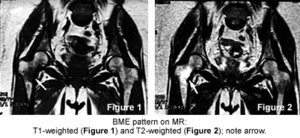
Bone marrow edema (BME) is a nonspecific finding associated with several different entities, including transient osteoporosis of the hip, transient bone-marrow edema syndrome, osteonecrosis, trauma, infection and infiltrative neoplasm. The BME pattern seen with MR consists of decreased T1-weighted and increased T2-weighted signal in the bone marrow and is consistent with increased free water or edema within normal fatty bone marrow.
This finding can be associated with several disease processes. Trauma, infection and neoplasm have clinical findings that allow us to differentiate them. However, osteonecrosis and the bone marrow edema syndromes' transient osteoporosis entities are not so easily differentiated. It is crucial that they be diagnosed correctly as they require very different treatment and intervention. It is the confusing terminology and diagnosis of bone marrow edema syndrome (BMES), transient regional osteoporosis and osteonecrosis that I will discuss in this brief article.
First, to review the terminology, there are several conditions that demonstrate the BME pattern: bone marrow edema syndrome (BMES), transient regional osteoporosis, transient osteoporosis of the hip and osteonecrosis. The main objective is to differentiate self-limiting conditions from osteonecrosis, which requires aggressive intervention.
Transient osteoporosis was first described in 1959 by Curtiss and Kincaid1 after finding an association between hip pain in the third trimester of pregnancy and radiographic bone demineralization. Since then, unfortunately, this condition has been described by several different terms. The term transient osteoporosis of the hip, described by Lequesne in 1968,2 was a condition thought to be seen mainly in women in their third trimester of pregnancy. However, it was later found that 60 percent of the cases are in men in their fourth through seventh decades of life.
The term bone marrow edema syndrome was introduced to replace transient osteoporosis of the hip based on characteristic MRI findings. The joints in the lower extremity are affected most often in the hip, but it also has been reported in the knee. Regional migratory osteoporosis (or migratory BMES), first described by Duncan, et al., as a migratory osteolysis,3 is characterized as a migrating arthralgia of the weight-bearing joints of the lower extremities associated with a focal osteoporosis. The migration occurs from one articulation to another and is the feature that separates regional migratory osteoporosis from BMES involving the hip or knee alone.
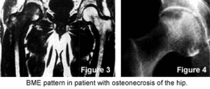
So, we have several terms that describe a process that may represent a spectrum of disease. Thus, depending on when the condition is first diagnosed, we may describe it with one term but later with another. Because they are all self-limiting conditions usually resolving within a two-year period, it may not matter which term is used, as long as it is differentiated from osteonecrosis (which is progressive and carries serious complications). Unfortunately, all these conditions present with bone marrow edema identified with the use of MRI, which is 100 percent sensitive but not specific. The edema seen with BMES usually is more extensive than that seen with osteonecrosis. The bone cortex may appear thinned but always is intact. Unlike osteonecosis, there should be no evidence of subchondral defects. Osteonecrosis of the hip is progressive, resulting from an interruption of the vascular supply to the femoral head and can lead to the collapse of the femoral head, destruction of the hip joint and necessitates aggressive intervention early in the disease process.
Early on, transient osteoporosis/BMES may be confused with osteonecrosis. In the past, there was much debate as to whether or not they are different degrees of the same disease. As it stands now, however, most clinicians are in agreement that transient osteoporosis/BMES does not lead to osteonecrosis. This is why differentiation between the self-limiting conditions and osteonecrosis is critical.
Transient osteoporosis/BMES is self-limiting; the natural history of this entity is that symptoms begin with no history of trauma and in-crease over a period of four to eight weeks, plateau, and then show a grad-ual resolution of symptoms over a three- to nine-month period. Migratory recurrence of symptoms usually can occur within the first two years after the initial pain relief.4
I am not aware of a chiropractic protocol for treating transient osteoporosis/BMES. Hopefully, this is only due to the fact that I have not done a sufficient literature search. The accepted medical treatment includes initiation of pain management and physical therapy with the use of protected weight-bearing. NSAIDs usually ameliorate the symptoms sufficiently for patients to manage their daily activities; however, stronger medication may be necessary. The main aim of the physical therapy is to prevent deconditioning,
which can lead to complications consisting of insufficiency fracture or subcapital fractures of the femoral neck that requires orthopedic intervention.
For this reason, protected weight-bearing is essential. Some recommend early introduction of weight-bearing activities, calcitonin, bisphosphonates or parathyroid hormone.5-9 Others have suggested that surgical core decompression may be used in BMES, especially in patients with refractory pain despite the use of conservative measures.10 Under certain circumstances, some cases need more intervention than others. I am not recommending any specific form of treatment. However, I believe chiropractors have been overlooked as appropriate providers of patients with transient osteoporosis/BMES, most likely due (in certain extent) to our lack of case reporting in peer-reviewed literature. As long as osteonecrosis has been ruled out, these conditions can be correctly identified and properly managed. Chiropractors are uniquely equipped to manage transient osteoporosis/BMES and should be identified as such.
References
Curtiss PH, Kincaid WE. Transitory demineralization of the hip in pregnancy: a report of three cases. J Bone Joint Surg Am, 1959;41:1327-33.
Lequesne M. Transient osteoporosis of the hip: a nontraumatic variety of Sudeck's atrophy. Ann Rheum Dis, 1968:27:463-71.
Duncan H, Frame B, Frost HM, Arnstein AR. Migratory oseolysis of the lower extremities. Ann Intern Med, 1967:66:165-73.
Lakhanpal S, Ginsburg WW, Luthra HS, Hunder GG. Transient regional osteoporosis: a study of 56 cases and review of the literature. Ann Intern Med, 1987;106:444-50.
Kibbi L, Touma Z, Khoury N, Arayassi T. Oral bisphosphonates in treatment of transient osteoporosis. Clin Rheumatol, 2007;14(34):9939.
Varenna M, et al. Intravenous pamidronate in the treatment of transient osteoporosis of the hip. Bone, 2002;31(1):96-101.
Schapira D, et al. Severe transient osteoporosis of the hip during pregnancy. Successful treatment with intravenous biphosphonates. Clin Exp Rheumatol, 2003;21(1):107-10.
Ringe JD, Dorst A, Faber H. Effective and rapid treatment of painful localized transient osteoporosis (bone-marrow edema) with intravenous ibandronate. Osteoporosis Int, 2005;16(12):2063-8.
Arayssi TK, et al. Calcitonin in the treatment of transient osteoporosis of the hip. Semin Arthritis Rheum, 2003;32(6):388-97.
Steinberg ME, Brighton CT, Steinberg DR, et al. Treatment of avascular necrosis of the femoral head by a combination of bone grafting, decompression and electrical stimulation. Clin Orthop Relat Res, 1984;186:137-53.
Many relevant diagnostic signs are not performed deliberately by the examiner or by the patient at the examiner’s direction. They are observed as the patient reacts to their condition. Fortin’s finger sign, Minor’s sign, and Vanzetti’s sign are three examples of this principle.
On Feb. 1, 2024, two more legislators – John R. Carter (R-Texas) and Robert “Bob” Good (R-Va.) – co-sponsored the Chiropractic Medicare Coverage Modernization Act of 2023, bringing the co-sponsor total to 155, two more than the total achieved over the entire two-year congressional cycle in which identical legislation last appeared (2021-22).
While working at the Lovelace Medical Center in Albuquerque, N.M., one of the primary care physicians stated that imaging every patient prior to receiving chiropractic was not medically necessary. It was at that point I reconsidered the need for the imaging of every patient to avoid malpractice.