Degenerative changes of the sacroiliac joint can be demonstrated pathologically in patients under 30 years of age. These generally are subtle alterations, and generally not detectable on radiographs. After the age of 40 years most patients reveal minimal signs of degenerative joint disease. The most common finding is decrease in joint space, which may involve the entire joint or focal areas of decreased joint space, mainly the inferior aspect. The interosseous space between the sacrum and ilium is normally 2 to 5 mm wide, reflecting the combined thickness of the sacral and iliac cartilage. With degenerative joint disease, pathologic abnormalities become prominent, particularly on the iliac side of the joint. Cartilage degeneration, subchondral eburnation and osteophytes become apparent; changes can be unilateral or bilateral and associated with significant clinical findings, including pain and tenderness.
Radiographic features associated with degenerative joint disease of the sacroiliac joint include: joint space loss, subchondral sclerosis and osteophytosis. With significant loss of subchondral bone of the ilium the joint may appear ankylosed with the sacrum. However, with close inspection of the trabeculae, one should be able to discern the lack of continuation of the trabeculae across the joint, which confirms the absence of ankylosis.
Osteophytosis is a prominent radiographic and pathologic characteristic of degenerative joint disease of the sacroiliac joint. It is more frequent in men than in women. Osteophytes can occur at any level in the joint but are most characteristic at the anterosuperior and anteroinferior margins of the joint. They may produce prominent focal radiopaque lesions, which can simulate the appearance of osteoblastic skeletal metastasis. With further growth of the osteophyte can extend across the anterior aspect of the articular cavity, causing periarticular bony ankylosis. This can simulate intra-articular bony ankylosis and can at times be falsely interpreted as ankylosing spondylitis. Fibrous ankylosis within the joint is typical of advanced degenerative joint disease of the sacroiliac joints; intra-articular bony ankylosis is exceedingly rare. This finding is generally associated with ankylosing spondylitis or one of the inflammatory arthropathies.
FIGURE 1: A spot on the sacrum demonstrates a radiopaque density overlying the left sacroiliac joint.
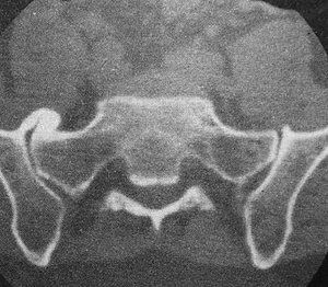
FIGURE 2: A large osteophytic formation is bridging the sacroiliac joint on one side, which on the plain film appears as a radiopaque density overlying the sacroiliac joint.
The figures are of a patient who was a 42-year-old male with a six year history of intermittent right-sided sacroiliac pain. The patient noted an incident of acute trauma: falling off a six foot sea wall, landing on his buttock and right hip. There was no apparent fracture to the hip or pelvis at the time of the incident. However, he experienced significant right hip and sacroiliac pain for several weeks, which gradually resolved, although three to four years later he developed intermittent right-sided sacroiliac pain.
Many relevant diagnostic signs are not performed deliberately by the examiner or by the patient at the examiner’s direction. They are observed as the patient reacts to their condition. Fortin’s finger sign, Minor’s sign, and Vanzetti’s sign are three examples of this principle.
Spearheaded by burgeoning scientific and clinical research literature, psychedelics have reached a level of media coverage and popular interest that has not been seen for over half a century. By “psychedelics,” we are referring to the unique class of substances that includes psilocybin (the active compound found in so-called “magic mushrooms”), LSD, dimethyltryptamine (DMT), ayahuasca, 5-MeO-DMT, and mescaline – each of which occurs in the natural world (except for LSD, which is a semi-synthetic compound).
While working at the Lovelace Medical Center in Albuquerque, N.M., one of the primary care physicians stated that imaging every patient prior to receiving chiropractic was not medically necessary. It was at that point I reconsidered the need for the imaging of every patient to avoid malpractice.