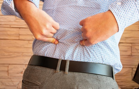
Many relevant diagnostic signs are not performed deliberately by the examiner or by the patient at the examiner’s direction. They are observed as the patient reacts to their condition. Fortin’s finger sign, Minor’s sign, and Vanzetti’s sign are three examples of this principle.
Do Injured Muscles Completely Repair?
All lacerations of muscles, whether partial or complete, heal with scar tissue (fibrosis). Even muscles that are injured in a nondisruptive manner (stretched to the plastic deformation level resulting in an alteration of their structure) show some regenerating muscle fibers, but normal histology is not restored and scar tissue is persistent.1
Garrett, et al.,2 state: "Data show that skeletal muscle can recover useful but not normal function after laceration and repair."
Muscles are able to regenerate after injury, so why do they often end up with incomplete functional recovery?
A recent article3 by Yong, Cummins and Huard in Current Opinion in Orthopaedics gives us some valuable information on this enigma. Whether it's due to direct trauma causing laceration and contusion, or indirect damage related to ischemia, strain or neurological dysfunction, the injured muscle initiates the regeneration of myofibers, but is "almost always accompanied by an overgrowth of fibroblast cells located within the connective tissue network." The initial injury damages the myofiber plasma membrane, resulting in ingress of calcium and a hematoma. Necrosis of the myofibers occurs; a hematoma may form within the injured muscle and help initiate an inflammatory process. Neutrophils appear within an hour, becoming a source of pro-inflammatory cytokines. Macrophages remain for several weeks. Lymphocytes debride the necrotic tissue and secrete a variety of growth factors that promote regeneration.
In simplistic form, the previous information represents the initial process of degeneration and inflammation. The next phase is regeneration, when satellite cells located between the sarcolemma and basement lamina of the muscle fibers are activated by growth factors, released by infiltrating mononuclear cells and platelets associated with the hematoma. The activated satellite cells proliferate and fuse into multinucleated myofibers.
For injured muscle to successfully repair, a delicate balance between muscle fiber regeneration and connective tissue growth must occur. But a pathological fibrosis occurs. The extracellular matrix (ECM), which is part of the connective tissue network found throughout skeletal muscle, is composed of fibroblastic cells and a complex mesh of several types of collagen, glycoproteins, and proteoglycans. The ECM causes an overproduction of several types of collagen, resulting in fibrosis. It has been found that the bone marrow is also stimulated during the injury process, and produces fibrotic-like cellsthat also contributing to the scar tissue (fibrosis).
"The formation of scar tissue not only blocks muscle regeneration but also leads to muscle that is far more susceptible to re-injury upon contraction."3 Yong, et al., found that transforming growth factor-B1 (TGF-B1) is a key factor in activating the fibrosis cascade in injured skeletal muscle.4 They have recently developed an antifibrosis agent called decorin, a proteoglycan that inactivates TGF-B1 and improves muscle healing to a near complete recovery. This substance is still in the experimental stage.
References
- Best TM, Garrett WE. Basic science of soft tissue: muscle and tendon. In: DeLee JC, Drez D. Orthopaedic Sports Medicine Principles & Practice, Vol 1, Philadelphia, WB Saunders, 1994.
- Garrett WE, Seaber AV, Boswick J, et al. Recovery of skeletal muscle after laceration and repair. J Hand Surg (Am) 1984 Sep;9(5):683-92.
- Yong LI, Cummins J, Huard J. Muscle injury and repair. Current Opinion in Orthopaedics 12(5), Oct. 2001:409-415.
- Li Y, Foster W, Sato K, et al. Non-viral mediated gene transfer of TGF-B1 promotes the differentiation of muscle derived cells toward fibroblastic lineage and muscle fibrosis. Mol Ther 2001,3:278.
Warren Hammer,MS,DC,DABCO
Norwalk, Connecticut
softissu@optonline.net


