Spina bifida occulta is often considered an incidental finding, and many times is not even mentioned in medical radiology reports. This vertebral anomaly is estimated to affect about one-fifth of the general population. There is, however, a higher incidence of this in populations with complaints of lower back pain.1 Spina bifida occulta was recognized as an embryologic developmental defect in 1642 by Nicholas Tult. There are several different pathophysiological mechanisms.
Spina bifida occulta can involve one or several adjacent vertebral segments, most commonly the transitional levels of the spine. It most often involves the first sacral segment, where it is associated with absence of the spinous process (SP) or lamina, or both. The unattached spinous process, if there is one, may remain in place, held by the ligamentum flavum, or it can fuse with the L5 SP to form a hooked or "tongue-shaped" process pointing toward the defect in the spinal canal. The L5 SP can also be unusually large (it is normally smaller than those of the other lumbar segments), and in the absence of the S1 SP, may protrude into the defect. All these situations are termed the "clasp-knife deformity," as described by Henry, et al.2
The intraspinal pathological changes associated with spina bifida occulta have been reported; upon surgery, the histologic abnormalities consist of fatty and fibrous tissue in contact with the nerves of the cauda equina, and often completely enveloping them. The dura is often incomplete or poorly formed at the level of the posterior arch defect. In extension, the L5 SP can encroach on the spinal canal.
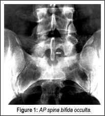
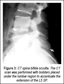
I will demonstrate one case where this was a definite factor in a patient's lower back pain. A 28-year-old male developed lower back pain at the age of 19 while lifting heavy boxes. He had intermittent recurrences of lower back pain since the initial episode. He described the pain as "pins and needles" in the base of his back. The pain radiated to the left hip, with associated intermittent numbness in the left calf. Symptoms were aggravated by standing and physical activity, and were relieved by lying down. He had no pain at night, but did complain of morning stiffness. Neurological examination revealed absent Babinski signs bilaterally, and diffuse, mild tenderness over the lower lumbar spinous processes and paraspinal muscles. Anterior-posterior (Figure 1) and lateral views (Figure 2) demonstrated spina bifida occulta at the S1 segment associated with an elongated L5 SP. Computerized tomography (CT) revealed spina bifida occulta, the classic clasp-knife deformity, with encroachment upon the spinal canal (Figure 3).
This patient responded to conservative treatment and was able to get almost total relief of symptoms, with only mild reoccurrences, generally caused by what the patient described as "just playing a good game of football or basketball."
My point in presenting this case is that often the lumbar region is not well visualized, and what we may feel is a classic presentation of disc disease may be something totally different. Not all patients with spina bifida occulta are symptomatic, but if this anomaly is present, the possibility should be explored.
References
Boone D, Parsons D, Lachmann SM, Sherwood T. Spina bifida occulta: Lesion or anomaly? Clinical Radiology 1985;36:159-161.
Henry GW, Larsen IJ, Stewart SF. The roentgenologic criteria for appraising the human back as an economic asset or liability. American Journal of Roentgenology 1938;39:937.
Many relevant diagnostic signs are not performed deliberately by the examiner or by the patient at the examiner’s direction. They are observed as the patient reacts to their condition. Fortin’s finger sign, Minor’s sign, and Vanzetti’s sign are three examples of this principle.
On Feb. 1, 2024, two more legislators – John R. Carter (R-Texas) and Robert “Bob” Good (R-Va.) – co-sponsored the Chiropractic Medicare Coverage Modernization Act of 2023, bringing the co-sponsor total to 155, two more than the total achieved over the entire two-year congressional cycle in which identical legislation last appeared (2021-22).
While working at the Lovelace Medical Center in Albuquerque, N.M., one of the primary care physicians stated that imaging every patient prior to receiving chiropractic was not medically necessary. It was at that point I reconsidered the need for the imaging of every patient to avoid malpractice.