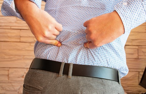
Many relevant diagnostic signs are not performed deliberately by the examiner or by the patient at the examiner’s direction. They are observed as the patient reacts to their condition. Fortin’s finger sign, Minor’s sign, and Vanzetti’s sign are three examples of this principle.
Posterior Spinal Fascial System
Every practitioner who treats spinal problems must become more aware of the fascial system, especially the fascial layers directly related to the spine. An article in the most recent issue of the American Journal of Sports Medicine1 describes an acute case of paraspinal muscle compartment syndrome. It stated that of three cases reported, none involved direct local trauma. While it was necessary to perform a fasciotomy, all were treated without surgery. The report stated that surgeons performed the operation because the myogenic enzyme value (creatine kinase) continued to rise.
In this case, physical findings showed a tender point at the left lumbar paraspinal muscle and decreased sensation in the left lumbosacral area. The recording of pressure of both paraspinal compartments were taken. Normal average should have been 4.3, and the abnormal left side was 14 to 16 mm Hg. After cutting the deep fascia, the superficial muscle layer appeared to be normal, but as they got to the deeper layer, the color changed from red to white. They ruled out a major contusion, since there was no hematoma in the muscle.
One day after the operation, the patient was able to walk, and the severe lower back pain abated. While we are familiar with a compartment syndrome occurring in the calf, due to restrictive fascia, we seldom think of a spinal problem in the same way. While the fascial spinal condition may not be as acute as a calf compartment, syndrome fascial restrictions often occur at paraspinal areas without reaching the compartment stage. The posterior layer in the lumbar region tightly encloses the paraspinal muscles and has been cited as a "rare" cause of exercise-induced low back pain.2
While a compartment syndrome may be rare regarding the lower back, tightened, shortened fascia based on its anatomy and function is undoubtedly related to spinal pain. In the young dancer, for example, the hyperlordosis is considered, due to a combination of muscle imbalance, relatively weak abdominal muscles and relatively tight lumbodorsal fascia.3
Another study proved that patients with chronic back pain versus patients without back pain had posterior fascia that was deficiently innervated.4 The posterior layer of spinal fascia is composed of a superficial and a deep layer which extends from the biceps femoris over the sacrotuberous ligament, up to the tendons of the splenius muscles superiorly. The posterior fascia facilitates the transfer of loads between the limbs and trunk.5 The deep layer blends anterolaterally with the transversus abdominus and internal oblique and the multifidus. The posterior fascia also attaches to the extrinsic back muscles such as the latissimus dorsi, gluteus medius and maximus. Vleeming, et al.,4 showed that traction to the latissimus dorsi created tension in the lumbar fascia which was transmitted to the gluteal muscles and biceps femoris. The posterior fascia acts as an intermediary in the transfer of loads between the upper and lower limbs, between the left and right sides of the body and between the abdominal walls and the spine.2 Since the posterior fascia tightly encloses the paraspinal muscles, it is thought that they produce a "hydraulic amplifier" effect by limiting radial expansion of the muscle during contraction, thereby increasing axial stress and improving muscle contraction.
Since the posterior fascia has viscoelastic properties, it alters its structure when exercised (and can also be altered by fascial release). Farfan thought that fascia thickened with exercise.6 This thickening can be beneficial by improving segmental stabilization, and load transfer capacity in the spine. Since load is transferred by way of the fascia from the gluteal muscles to the opposite latissimus dorsi, exercise such as swimming which involves contralateral muscle groups may be beneficial. The bracing effect of the thoracolumbar fascia on the lower lumbar spine and sacroiliac joints is essential for proper load transfer between the spine and lower extremities.
Fascia should not be too tight or too loose. Restricted fascia alters the transfer of load across the sacroiliac joint and anywhere else it transfers load, i.e., from the lower to the upper extremities; from the pelvis to the occiput; and from the anterolateral abdominals to the spine. Clearly, it is necessary to evaluate fascia for shortening. There is increased erector spinae pressure when a standing individual flexes forward.
A good test for tight posterior spinal fascia is to have a standing or sitting patient bend in forward flexion. Next, the patient bends forward in left or right rotation while the practitioner pushes up on the posterior shoulder to stretch the latissimus dorsi. Areas of abnormal tension or pain will appear, and will disappear with fascial release. Barker2 remarks that since fasciae transmit tension between the cervical and lumbar spine, in any whiplash-type injury the "variability in fascial thickness and strength might then account for variation in symptoms between individuals of differing builds."2 Another reason for variability in symptoms will be the chronic fascial restrictions that can no longer act as shock absorbers, thereby absorbing more stress in a local area.
References
- Izuru K, Tachibana S, Hirota Y, et al. Acute paraspinal muscle compartment syndrome treated with surgical decompression. Amer J Sports Med 2002;30(2):283-285.
- Barker PJ, Briggs CA. Attachments of the posterior layer of lumbar fascia. Spine 1999;2(17):1757-1764.
- Solomon R, Brown T, Gerbino C, Micheli LJ. The young dancer. Clin In Sports Med 2000;19(4):717-739.
- Yahia LH, Pigeon P, Des Rosiers EA. Viscoelastic properties of the human lumbodorsal fascia. J Biomed Eng 1993;15:425-429.
- Vleeming A, Pool-Goudzwaard AL, Stoekart T, et al. The posterior layer of the thoracolumbar fascia: Its function in load transfer from spine to legs. Spine 1995;21:753-8.
- Farfan HF. Form and function of the musculoskeletal system as revealed by mathematical analysis of the lumbar sine: An essay. Spine 1995;20:1462-74.
Warren Hammer,MS,DC,DABCO
Norwalk, Connecticut
softissu@optonline.net


